 Movie
Movie Controller


+ Open data

- Basic information
| Entry | Database: PDB / ID: 8isy | ||||||
|---|---|---|---|---|---|---|---|
| Title | Cryo-EM structure of free-state Crt-SPARTA | ||||||
 Components Components |
| ||||||
 Keywords Keywords |  DNA BINDING PROTEIN / Ago / DNA/RNA DNA BINDING PROTEIN / Ago / DNA/RNA | ||||||
| Function / homology | TIR domain / Toll/interleukin-1 receptor homology (TIR) domain / Toll/interleukin-1 receptor homology (TIR) domain superfamily / Ribonuclease H superfamily / nucleic acid binding / Ribonuclease H-like superfamily / signal transduction / Piwi domain-containing protein / TIR domain-containing protein Function and homology information Function and homology information | ||||||
| Biological species |  Thermoflavifilum thermophilum (bacteria) Thermoflavifilum thermophilum (bacteria) | ||||||
| Method | ELECTRON MICROSCOPY / single particle reconstruction / cryo EM / Resolution: 3.27 Å | ||||||
 Authors Authors | Gao, X. / Shang, K. / Zhu, K. / Wang, L. / Mu, Z. / Fu, X. / Yu, X. / Qin, B. / Zhu, H. / Ding, W. / Cui, S. | ||||||
| Funding support | 1items
| ||||||
 Citation Citation | Journal: Nature / Year: 2024 Title: Nucleic-acid-triggered NADase activation of a short prokaryotic Argonaute. Authors: Xiaopan Gao / Kun Shang / Kaixiang Zhu / Linyue Wang / Zhixia Mu / Xingke Fu / Xia Yu / Bo Qin / Hongtao Zhu / Wei Ding / Sheng Cui /  Abstract: Argonaute (Ago) proteins mediate RNA- or DNA-guided inhibition of nucleic acids. Although the mechanisms used by eukaryotic Ago proteins and long prokaryotic Ago proteins (pAgos) are known, that used ...Argonaute (Ago) proteins mediate RNA- or DNA-guided inhibition of nucleic acids. Although the mechanisms used by eukaryotic Ago proteins and long prokaryotic Ago proteins (pAgos) are known, that used by short pAgos remains elusive. Here we determined the cryo-electron microscopy structures of a short pAgo and the associated TIR-APAZ proteins (SPARTA) from Crenotalea thermophila (Crt): a free-state Crt-SPARTA; a guide RNA-target DNA-loaded Crt-SPARTA; two Crt-SPARTA dimers with distinct TIR organization; and a Crt-SPARTA tetramer. These structures reveal that Crt-SPARTA is composed of a bilobal-fold Ago lobe that connects with a TIR lobe. Whereas the Crt-Ago contains a MID and a PIWI domain, Crt-TIR-APAZ has a TIR domain, an N-like domain, a linker domain and a trigger domain. The bound RNA-DNA duplex adopts a B-form conformation that is recognized by base-specific contacts. Nucleic acid binding causes conformational changes because the trigger domain acts as a 'roadblock' that prevents the guide RNA 5' ends and the target DNA 3' ends from reaching their canonical pockets; this disorders the MID domain and promotes Crt-SPARTA dimerization. Two RNA-DNA-loaded Crt-SPARTA dimers form a tetramer through their TIR domains. Four Crt-TIR domains assemble into two parallel head-to-tail-organized TIR dimers, indicating an NADase-active conformation, which is supported by our mutagenesis study. Our results reveal the structural basis of short-pAgo-mediated defence against invading nucleic acids, and provide insights for optimizing the detection of SPARTA-based programmable DNA sequences. | ||||||
| History |
|
- Structure visualization
| Structure viewer | Molecule: MolmilJmol/JSmol |
|---|
- Downloads & links
-Download
| PDBx/mmCIF format | 8isy.cif.gz | 198.9 KB | Display | PDBx/mmCIF format |
|---|---|---|---|---|
| PDB format | pdb8isy.ent.gz | 156.1 KB | Display | PDB format |
| PDBx/mmJSON format | 8isy.json.gz | Tree view | PDBx/mmJSON format | |
| Others |  Other downloads Other downloads |
-Validation report
| Arichive directory | https://data.pdbj.org/pub/pdb/validation_reports/is/8isyftp://data.pdbj.org/pub/pdb/validation_reports/is/8isy | HTTPS FTP |
|---|
-Related structure data
| Related structure data |  35700MC 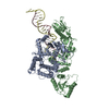 8iszC 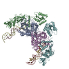 8it0C 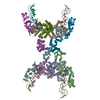 8it1C 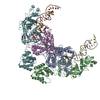 8k9gC M: map data used to model this data C: citing same article ( |
|---|---|
| Similar structure data |


-Links
 PDBj
PDBj
- Assembly
| Deposited unit | 
|
|---|---|
| 1 |
|
-Components
| #1: Protein | Mass: 58304.848 Da / Num. of mol.: 1 Source method: isolated from a genetically manipulated source Source: (gene. exp.) Thermoflavifilum thermophilum (bacteria)Gene: SAMN05660895_1671 / Production host: Expression vector pET-mod (others) / References: UniProt: A0A1I7NFD7 |
|---|---|
| #2: Protein | Mass: 53256.734 Da / Num. of mol.: 1 Source method: isolated from a genetically manipulated source Source: (gene. exp.) Thermoflavifilum thermophilum (bacteria)Gene: SAMN05660895_1670 / Production host: Expression vector pET-mod (others) / References: UniProt: A0A1I7NFG5 |
-Experimental details
-Experiment
| Experiment | Method: ELECTRON MICROSCOPY |
|---|---|
| EM experiment | Aggregation state: PARTICLE / 3D reconstruction method: single particle reconstruction |
- Sample preparation
| Component | Name: prokaryotic Argonaute System / Type: COMPLEX / Entity ID: all / Source: RECOMBINANT |
|---|---|
| Source (natural) | Organism: Thermoflavifilum thermophilum (bacteria) |
| Source (recombinant) | Organism: Expression vector pET-mod (others) |
| Buffer solution | pH: 8 |
| Specimen | Embedding applied: NO / Shadowing applied: NO / Staining applied: NO / Vitrification applied: YES |
| Specimen support | Grid material: GOLD / Grid type: Quantifoil R1.2/1.3 |
| Vitrification | Cryogen name: ETHANE |
- Electron microscopy imaging
| Experimental equipment |  Model: Titan Krios / Image courtesy: FEI Company |
|---|---|
| Microscopy | Model: FEI TITAN KRIOS |
| Electron gun | Electron source: FIELD EMISSION GUN / Accelerating voltage: 300 kV / Illumination mode: FLOOD BEAM |
| Electron lens | Mode: BRIGHT FIELDBright-field microscopy / Nominal defocus max: 2500 nm / Nominal defocus min: 1800 nm / Calibrated defocus min: 1800 nm / Calibrated defocus max: 2500 nm / Alignment procedure: COMA FREE |
| Specimen holder | Cryogen: NITROGEN / Specimen holder model: FEI TITAN KRIOS AUTOGRID HOLDER |
| Image recording | Electron dose: 66 e/Å2 / Film or detector model: GATAN K3 (6k x 4k) |
- Processing
| EM software |
| ||||||||||||||||||||||||
|---|---|---|---|---|---|---|---|---|---|---|---|---|---|---|---|---|---|---|---|---|---|---|---|---|---|
| CTF correction | Type: PHASE FLIPPING AND AMPLITUDE CORRECTION | ||||||||||||||||||||||||
| Symmetry | Point symmetry: C1 (asymmetric) | ||||||||||||||||||||||||
| 3D reconstruction | Resolution: 3.27 Å / Resolution method: FSC 0.143 CUT-OFF / Num. of particles: 1158576 / Symmetry type: POINT | ||||||||||||||||||||||||
| Atomic model building | Protocol: FLEXIBLE FIT / Space: REAL / Target criteria: Correlation coefficient | ||||||||||||||||||||||||
| Refine LS restraints |
|
