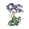
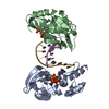
 Movie
Movie Controller


+ Open data

- Basic information
| Entry | Database: PDB / ID: 8ilg | |||||||||||||||
|---|---|---|---|---|---|---|---|---|---|---|---|---|---|---|---|---|
| Title | The crystal structure of dG-DNA:Pol X product binary complex | |||||||||||||||
 Components Components |
| |||||||||||||||
 Keywords Keywords | REPLICATION/DNA /  DNA polymerase / substrate complex / selenium-modified dNTP / configuration / REPLICATION-DNA complex DNA polymerase / substrate complex / selenium-modified dNTP / configuration / REPLICATION-DNA complex | |||||||||||||||
| Function / homology |  Function and homology informationvirion component / base-excision repair / double-strand break repair via nonhomologous end joining / DNA-directed DNA polymerase / DNA-directed DNA polymerase activity / DNA binding / metal ion binding Function and homology informationvirion component / base-excision repair / double-strand break repair via nonhomologous end joining / DNA-directed DNA polymerase / DNA-directed DNA polymerase activity / DNA binding / metal ion bindingSimilarity search - Function | |||||||||||||||
| Biological species |   African swine fever virus African swine fever virussynthetic construct (others) | |||||||||||||||
| Method | X-RAY DIFFRACTION / SYNCHROTRON / MOLECULAR REPLACEMENT / Resolution: 1.805 Å | |||||||||||||||
 Authors Authors | Qin, T. / Gan, J.H. / Huang, Z. | |||||||||||||||
| Funding support |  China, 4items China, 4items
| |||||||||||||||
 Citation Citation | Journal: Int J Mol Sci / Year: 2023 Title: Structural Insight into Polymerase Mechanism via a Chiral Center Generated with a Single Selenium Atom. Authors: Qin, T. / Hu, B. / Zhao, Q. / Wang, Y. / Wang, S. / Luo, D. / Lyu, J. / Chen, Y. / Gan, J. / Huang, Z. | |||||||||||||||
| History |
|
- Structure visualization
| Structure viewer | Molecule: MolmilJmol/JSmol |
|---|
- Downloads & links
-Download
| PDBx/mmCIF format | 8ilg.cif.gz | 213.2 KB | Display | PDBx/mmCIF format |
|---|---|---|---|---|
| PDB format | pdb8ilg.ent.gz | 165.6 KB | Display | PDB format |
| PDBx/mmJSON format | 8ilg.json.gz | Tree view | PDBx/mmJSON format | |
| Others |  Other downloads Other downloads |
-Validation report
| Arichive directory | https://data.pdbj.org/pub/pdb/validation_reports/il/8ilgftp://data.pdbj.org/pub/pdb/validation_reports/il/8ilg | HTTPS FTP |
|---|
-Related structure data


-Links
 PDBj
PDBj

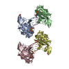
- Assembly
| Deposited unit | 
| ||||||||
|---|---|---|---|---|---|---|---|---|---|
| 1 |
| ||||||||
| 2 |
| ||||||||
| Unit cell |
|
-Components
| #1: Protein | Mass: 20536.668 Da / Num. of mol.: 2 Source method: isolated from a genetically manipulated source Source: (gene. exp.) African swine fever virus (strain Badajoz 1971 Vero-adapted)Gene: Ba71V-97, O174L / Production host:  Escherichia coli (E. coli) / References: UniProt: P42494, DNA-directed DNA polymerase Escherichia coli (E. coli) / References: UniProt: P42494, DNA-directed DNA polymerase#2: DNA chain | Mass: 2427.605 Da / Num. of mol.: 4 / Source method: obtained synthetically / Source: (synth.) synthetic construct (others) #3: Chemical |   Mass: 54.938 Da / Num. of mol.: 2 / Source method: obtained synthetically / Formula: Mn / Feature type: SUBJECT OF INVESTIGATION Mass: 54.938 Da / Num. of mol.: 2 / Source method: obtained synthetically / Formula: Mn / Feature type: SUBJECT OF INVESTIGATION#4: Chemical | Formic acid  Mass: 46.025 Da / Num. of mol.: 2 / Source method: obtained synthetically / Formula: CH2O2 Mass: 46.025 Da / Num. of mol.: 2 / Source method: obtained synthetically / Formula: CH2O2#5: Water | ChemComp-HOH / | Water Mass: 18.015 Da / Num. of mol.: 537 / Source method: isolated from a natural source / Formula: H2O Mass: 18.015 Da / Num. of mol.: 537 / Source method: isolated from a natural source / Formula: H2OHas ligand of interest | Y | |
|---|
-Experimental details
-Experiment
| Experiment | Method: X-RAY DIFFRACTION / Number of used crystals: 1 |
|---|
- Sample preparation
| Crystal | Density Matthews: 2.57 Å3/Da / Density % sol: 52.06 % |
|---|---|
| Crystal grow | Temperature: 289.15 K / Method: vapor diffusion, hanging drop / Details: Potassium Formate (0.2 M), 16% PEG3350 |
-Data collection
| Diffraction | Mean temperature: 100 K / Serial crystal experiment: N | |||||||||||||||||||||||||||||||||||||||||||||||||||||||||||||||||||||||||||||
|---|---|---|---|---|---|---|---|---|---|---|---|---|---|---|---|---|---|---|---|---|---|---|---|---|---|---|---|---|---|---|---|---|---|---|---|---|---|---|---|---|---|---|---|---|---|---|---|---|---|---|---|---|---|---|---|---|---|---|---|---|---|---|---|---|---|---|---|---|---|---|---|---|---|---|---|---|---|---|
| Diffraction source | Source: SYNCHROTRON / Site: SSRF / Beamline: BL18U1 / Wavelength: 0.979 Å | |||||||||||||||||||||||||||||||||||||||||||||||||||||||||||||||||||||||||||||
| Detector | Type: MARMOSAIC 325 mm CCD / Detector: CCD / Date: May 7, 2020 | |||||||||||||||||||||||||||||||||||||||||||||||||||||||||||||||||||||||||||||
| Radiation | Protocol: SINGLE WAVELENGTH / Monochromatic (M) / Laue (L): M / Scattering type: x-ray | |||||||||||||||||||||||||||||||||||||||||||||||||||||||||||||||||||||||||||||
| Radiation wavelength | Wavelength: 0.979 Å / Relative weight: 1 | |||||||||||||||||||||||||||||||||||||||||||||||||||||||||||||||||||||||||||||
| Reflection | Resolution: 1.8→30 Å / Num. obs: 84882 / % possible obs: 91.6 % / Redundancy: 2.5 % / Rmerge(I) obs: 0.136 / Χ2: 0.111 / Net I/σ(I): 6.1 / Num. measured all: 209940 | |||||||||||||||||||||||||||||||||||||||||||||||||||||||||||||||||||||||||||||
| Reflection shell |
|
- Processing
| Software |
| ||||||||||||||||||||||||||||||||||||||||||||||||||||||||||||||||||||||||||||||||||||||||||||||||||||||||||||||||||||||||||||||
|---|---|---|---|---|---|---|---|---|---|---|---|---|---|---|---|---|---|---|---|---|---|---|---|---|---|---|---|---|---|---|---|---|---|---|---|---|---|---|---|---|---|---|---|---|---|---|---|---|---|---|---|---|---|---|---|---|---|---|---|---|---|---|---|---|---|---|---|---|---|---|---|---|---|---|---|---|---|---|---|---|---|---|---|---|---|---|---|---|---|---|---|---|---|---|---|---|---|---|---|---|---|---|---|---|---|---|---|---|---|---|---|---|---|---|---|---|---|---|---|---|---|---|---|---|---|---|---|
| Refinement | Method to determine structure: MOLECULAR REPLACEMENT / Resolution: 1.805→28.861 Å / SU ML: 0.23 / Cross valid method: FREE R-VALUE / σ(F): 1.37 / Phase error: 28.42 / Stereochemistry target values: ML
| ||||||||||||||||||||||||||||||||||||||||||||||||||||||||||||||||||||||||||||||||||||||||||||||||||||||||||||||||||||||||||||||
| Solvent computation | Shrinkage radii: 0.9 Å / VDW probe radii: 1.11 Å / Solvent model: FLAT BULK SOLVENT MODEL | ||||||||||||||||||||||||||||||||||||||||||||||||||||||||||||||||||||||||||||||||||||||||||||||||||||||||||||||||||||||||||||||
| Refinement step | Cycle: LAST / Resolution: 1.805→28.861 Å
| ||||||||||||||||||||||||||||||||||||||||||||||||||||||||||||||||||||||||||||||||||||||||||||||||||||||||||||||||||||||||||||||
| Refine LS restraints |
| ||||||||||||||||||||||||||||||||||||||||||||||||||||||||||||||||||||||||||||||||||||||||||||||||||||||||||||||||||||||||||||||
| LS refinement shell |
| ||||||||||||||||||||||||||||||||||||||||||||||||||||||||||||||||||||||||||||||||||||||||||||||||||||||||||||||||||||||||||||||
| Refinement TLS params. | Method: refined / Origin x: 87.1167 Å / Origin y: 30.2348 Å / Origin z: 115.6841 Å
| ||||||||||||||||||||||||||||||||||||||||||||||||||||||||||||||||||||||||||||||||||||||||||||||||||||||||||||||||||||||||||||||
| Refinement TLS group | Selection details: all |
