 Movie
Movie Controller

[English] 日本語
 Yorodumi
- PDB-8il8: Crystal structure of Pyruvic Oxime Dioxygenase (POD) from Alcalig... -
+ Open data

- Basic information
| Entry | Database: PDB / ID: 8il8 | |||||||||
|---|---|---|---|---|---|---|---|---|---|---|
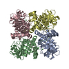
| Title | Crystal structure of Pyruvic Oxime Dioxygenase (POD) from Alcaligenes faecalis | |||||||||
 Components Components | Aldolase Fructose-bisphosphate aldolase Fructose-bisphosphate aldolase | |||||||||
 Keywords Keywords | OXIDOREDUCTASE / class II aldolase-like / dioxygenase / non-heme iron / His-triad | |||||||||
| Function / homology | Class II aldolase/adducin N-terminal / Class II Aldolase and Adducin N-terminal domain / Class II Aldolase and Adducin N-terminal domain / Class II aldolase/adducin N-terminal domain superfamily / : / Aldolase Function and homology information Function and homology information | |||||||||
| Biological species |  Alcaligenes faecalis subsp. faecalis NBRC 13111 (bacteria) Alcaligenes faecalis subsp. faecalis NBRC 13111 (bacteria) | |||||||||
| Method | X-RAY DIFFRACTION / SYNCHROTRON / MOLECULAR REPLACEMENT / Resolution: 1.769 Å | |||||||||
 Authors Authors | Tsujino, S. / Yamada, Y. / Fujiwara, T. | |||||||||
| Funding support |  Japan, 2items Japan, 2items
| |||||||||
 Citation Citation | Journal: To Be Published Title: Structural and functional analysis of pyruvic oxime dioxygenase, a key enzyme of heterotrophic nitrification Authors: Tsujino, S. / Yamada, Y. / Senda, M. / Senda, T. / Fujiwara, T. | |||||||||
| History |
|
- Structure visualization
| Structure viewer | Molecule: MolmilJmol/JSmol |
|---|
- Downloads & links
-Download
| PDBx/mmCIF format | 8il8.cif.gz | 372.4 KB | Display | PDBx/mmCIF format |
|---|---|---|---|---|
| PDB format | pdb8il8.ent.gz | 303.8 KB | Display | PDB format |
| PDBx/mmJSON format | 8il8.json.gz | Tree view | PDBx/mmJSON format | |
| Others |  Other downloads Other downloads |
-Validation report
| Arichive directory | https://data.pdbj.org/pub/pdb/validation_reports/il/8il8ftp://data.pdbj.org/pub/pdb/validation_reports/il/8il8 | HTTPS FTP |
|---|
-Related structure data

-Links
 PDBj
PDBj


- Assembly
| Deposited unit | 
| ||||||||
|---|---|---|---|---|---|---|---|---|---|
| 1 |
| ||||||||
| Unit cell |
|
-Components
| #1: Protein | Fructose-bisphosphate aldolase / Pyruvic oxime dioxygenase Mass: 29138.920 Da / Num. of mol.: 4 Source method: isolated from a genetically manipulated source Source: (gene. exp.) Alcaligenes faecalis subsp. faecalis NBRC 13111 (bacteria)Production host: Escherichia coli BL21(DE3) (bacteria) / References: UniProt: A0A0A2N3A3#2: Chemical | ChemComp-FE2 /   Mass: 55.845 Da / Num. of mol.: 4 / Source method: obtained synthetically / Formula: Fe / Feature type: SUBJECT OF INVESTIGATION Mass: 55.845 Da / Num. of mol.: 4 / Source method: obtained synthetically / Formula: Fe / Feature type: SUBJECT OF INVESTIGATION#3: Water | ChemComp-HOH / | Water Mass: 18.015 Da / Num. of mol.: 693 / Source method: isolated from a natural source / Formula: H2O Mass: 18.015 Da / Num. of mol.: 693 / Source method: isolated from a natural source / Formula: H2OHas ligand of interest | Y | |
|---|
-Experimental details
-Experiment
| Experiment | Method: X-RAY DIFFRACTION / Number of used crystals: 1 |
|---|
- Sample preparation
| Crystal | Density Matthews: 2.2 Å3/Da / Density % sol: 44.1 % |
|---|---|
| Crystal grow | Temperature: 293 K / Method: vapor diffusion, sitting drop / pH: 7.5 Details: 36% (v/v) PEG 400, 0.2M calcium acetate, 0.1M HEPES |
-Data collection
| Diffraction | Mean temperature: 95 K / Serial crystal experiment: N |
|---|---|
| Diffraction source | Source: SYNCHROTRON / Site: Photon Factory / Beamline: BL-17A / Wavelength: 0.98 Å |
| Detector | Type: DECTRIS EIGER X 16M / Detector: PIXEL / Date: Jun 7, 2022 |
| Radiation | Protocol: SINGLE WAVELENGTH / Monochromatic (M) / Laue (L): M / Scattering type: x-ray |
| Radiation wavelength | Wavelength: 0.98 Å / Relative weight: 1 |
| Reflection | Resolution: 1.76→76.44 Å / Num. obs: 55281 / % possible obs: 93.5 % / Redundancy: 3.4 % / Rmerge(I) obs: 0.056 / Net I/σ(I): 8.9 |
| Reflection shell | Resolution: 1.76→1.9 Å / Rmerge(I) obs: 0.565 / Num. unique obs: 2766 |
- Processing
| Software |
| |||||||||||||||||||||||||||||||||||||||||||||||||||||||||||||||||||||||||||||||||||||||||||||||||||||||||||||||||||||||||||||
|---|---|---|---|---|---|---|---|---|---|---|---|---|---|---|---|---|---|---|---|---|---|---|---|---|---|---|---|---|---|---|---|---|---|---|---|---|---|---|---|---|---|---|---|---|---|---|---|---|---|---|---|---|---|---|---|---|---|---|---|---|---|---|---|---|---|---|---|---|---|---|---|---|---|---|---|---|---|---|---|---|---|---|---|---|---|---|---|---|---|---|---|---|---|---|---|---|---|---|---|---|---|---|---|---|---|---|---|---|---|---|---|---|---|---|---|---|---|---|---|---|---|---|---|---|---|---|
| Refinement | Method to determine structure: MOLECULAR REPLACEMENT / Resolution: 1.769→76.44 Å / Cor.coef. Fo:Fc: 0.943 / Cor.coef. Fo:Fc free: 0.935 / SU R Cruickshank DPI: 0.222 / Cross valid method: THROUGHOUT / SU R Blow DPI: 0.233 / SU Rfree Blow DPI: 0.172 / SU Rfree Cruickshank DPI: 0.171
| |||||||||||||||||||||||||||||||||||||||||||||||||||||||||||||||||||||||||||||||||||||||||||||||||||||||||||||||||||||||||||||
| Displacement parameters | Biso mean: 32.73 Å2
| |||||||||||||||||||||||||||||||||||||||||||||||||||||||||||||||||||||||||||||||||||||||||||||||||||||||||||||||||||||||||||||
| Refine analyze | Luzzati coordinate error obs: 0.24 Å | |||||||||||||||||||||||||||||||||||||||||||||||||||||||||||||||||||||||||||||||||||||||||||||||||||||||||||||||||||||||||||||
| Refinement step | Cycle: LAST / Resolution: 1.769→76.44 Å
| |||||||||||||||||||||||||||||||||||||||||||||||||||||||||||||||||||||||||||||||||||||||||||||||||||||||||||||||||||||||||||||
| Refine LS restraints |
| |||||||||||||||||||||||||||||||||||||||||||||||||||||||||||||||||||||||||||||||||||||||||||||||||||||||||||||||||||||||||||||
| LS refinement shell | Resolution: 1.77→1.84 Å
| |||||||||||||||||||||||||||||||||||||||||||||||||||||||||||||||||||||||||||||||||||||||||||||||||||||||||||||||||||||||||||||
| Refinement TLS params. | Refine-ID: X-RAY DIFFRACTION
| |||||||||||||||||||||||||||||||||||||||||||||||||||||||||||||||||||||||||||||||||||||||||||||||||||||||||||||||||||||||||||||
| Refinement TLS group |
|
