 Movie
Movie Controller


+ Open data

- Basic information
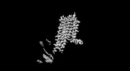
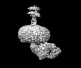
| Entry | Database: PDB / ID: 8ikl | ||||||
|---|---|---|---|---|---|---|---|
| Title | Cryo-EM structure of the CD97-G13 complex | ||||||
 Components Components |
| ||||||
 Keywords Keywords |  MEMBRANE PROTEIN / adhension GPCR / CD97 / G13 / complex MEMBRANE PROTEIN / adhension GPCR / CD97 / G13 / complex | ||||||
| Function / homology |  Function and homology information Function and homology informationClass B/2 (Secretin family receptors) / secretory granule membrane / G protein-coupled receptor activity / Olfactory Signaling Pathway / Activation of the phototransduction cascade / G beta:gamma signalling through PLC beta / Presynaptic function of Kainate receptors / Thromboxane signalling through TP receptor / G-protein activation / G protein-coupled acetylcholine receptor signaling pathway ...Class B/2 (Secretin family receptors) / secretory granule membrane / G protein-coupled receptor activity / Olfactory Signaling Pathway / Activation of the phototransduction cascade / G beta:gamma signalling through PLC beta / Presynaptic function of Kainate receptors / Thromboxane signalling through TP receptor / G-protein activation / G protein-coupled acetylcholine receptor signaling pathway / Activation of G protein gated Potassium channels / Inhibition of voltage gated Ca2+ channels via Gbeta/gamma subunits / Prostacyclin signalling through prostacyclin receptor / Glucagon signaling in metabolic regulation / G beta:gamma signalling through CDC42 / adenylate cyclase-activating G protein-coupled receptor signaling pathway / ADP signalling through P2Y purinoceptor 12 / G beta:gamma signalling through BTK / Sensory perception of sweet, bitter, and umami (glutamate) taste / Synthesis, secretion, and inactivation of Glucagon-like Peptide-1 (GLP-1) / photoreceptor disc membrane / Adrenaline,noradrenaline inhibits insulin secretion / Glucagon-type ligand receptors / Vasopressin regulates renal water homeostasis via Aquaporins / G alpha (z) signalling events / cellular response to catecholamine stimulus / Glucagon-like Peptide-1 (GLP1) regulates insulin secretion / ADORA2B mediated anti-inflammatory cytokines production / adenylate cyclase-activating dopamine receptor signaling pathway / ADP signalling through P2Y purinoceptor 1 / G beta:gamma signalling through PI3Kgamma / cellular response to prostaglandin E stimulus / Cooperation of PDCL (PhLP1) and TRiC/CCT in G-protein beta folding / sensory perception of taste / GPER1 signaling / G-protein beta-subunit binding / heterotrimeric G-protein complex / Inactivation, recovery and regulation of the phototransduction cascade / extracellular vesicle / G alpha (12/13) signalling events / transmembrane signaling receptor activity / signaling receptor complex adaptor activity / Thrombin signalling through proteinase activated receptors (PARs) / retina development in camera-type eye / cell-cell signaling / GTPase binding / Ca2+ pathway / phospholipase C-activating G protein-coupled receptor signaling pathway / G alpha (i) signalling events / fibroblast proliferation / G alpha (s) signalling events / G alpha (q) signalling events / Ras protein signal transduction / cell population proliferation / Extra-nuclear estrogen signaling / cell surface receptor signaling pathway / cell adhesion / immune response / inflammatory response / G protein-coupled receptor signaling pathway / lysosomal membrane / focal adhesion / GTPase activity / synapse / calcium ion binding / Neutrophil degranulation / protein-containing complex binding / signal transduction / extracellular exosome / membrane / plasma membrane / cytosol / cytoplasmSimilarity search - Function | ||||||
| Biological species |  Homo sapiens (human) Homo sapiens (human) | ||||||
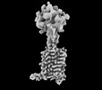
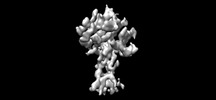
| Method | ELECTRON MICROSCOPY / single particle reconstruction / cryo EM / Resolution: 2.33 Å | ||||||
 Authors Authors | Mao, C. / Zhao, R. / Dong, Y. / Gao, M. / Chen, L. / Zhang, C. / Xiao, P. | ||||||
| Funding support |  China, 1items China, 1items
| ||||||
 Citation Citation | Journal: Mol Cell / Year: 2024 Title: Conformational transitions and activation of the adhesion receptor CD97. Authors: Chunyou Mao / Ru-Jia Zhao / Ying-Jun Dong / Mingxin Gao / Li-Nan Chen / Chao Zhang / Peng Xiao / Jia Guo / Jiao Qin / Dan-Dan Shen / Su-Yu Ji / Shao-Kun Zang / Huibing Zhang / Wei-Wei Wang / ...Authors: Chunyou Mao / Ru-Jia Zhao / Ying-Jun Dong / Mingxin Gao / Li-Nan Chen / Chao Zhang / Peng Xiao / Jia Guo / Jiao Qin / Dan-Dan Shen / Su-Yu Ji / Shao-Kun Zang / Huibing Zhang / Wei-Wei Wang / Qingya Shen / Jin-Peng Sun / Yan Zhang / Abstract: Adhesion G protein-coupled receptors (aGPCRs) are evolutionarily ancient receptors involved in a variety of physiological and pathophysiological processes. Modulators of aGPCR, particularly ...Adhesion G protein-coupled receptors (aGPCRs) are evolutionarily ancient receptors involved in a variety of physiological and pathophysiological processes. Modulators of aGPCR, particularly antagonists, hold therapeutic promise for diseases like cancer and immune and neurological disorders. Hindered by the inactive state structural information, our understanding of antagonist development and aGPCR activation faces challenges. Here, we report the cryo-electron microscopy structures of human CD97, a prototypical aGPCR that plays crucial roles in immune system, in its inactive apo and G13-bound fully active states. Compared with other family GPCRs, CD97 adopts a compact inactive conformation with a constrained ligand pocket. Activation induces significant conformational changes for both extracellular and intracellular sides, creating larger cavities for Stachel sequence binding and G13 engagement. Integrated with functional and metadynamics analyses, our study provides significant mechanistic insights into the activation and signaling of aGPCRs, paving the way for future drug discovery efforts. | ||||||
| History |
|
- Structure visualization
| Structure viewer | Molecule: MolmilJmol/JSmol |
|---|
- Downloads & links
-Download
| PDBx/mmCIF format | 8ikl.cif.gz | 164.5 KB | Display | PDBx/mmCIF format |
|---|---|---|---|---|
| PDB format | pdb8ikl.ent.gz | 126.2 KB | Display | PDB format |
| PDBx/mmJSON format | 8ikl.json.gz | Tree view | PDBx/mmJSON format | |
| Others |  Other downloads Other downloads |
-Validation report
| Arichive directory | https://data.pdbj.org/pub/pdb/validation_reports/ik/8iklftp://data.pdbj.org/pub/pdb/validation_reports/ik/8ikl | HTTPS FTP |
|---|
-Related structure data
| Related structure data |  35516MC 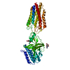 8ikjC M: map data used to model this data C: citing same article ( |
|---|---|
| Similar structure data |
-Links
 PDBj
PDBj


- Assembly
| Deposited unit | 
|
|---|---|
| 1 |
|
-Components
| #1: Protein | Mass: 31429.586 Da / Num. of mol.: 1 Source method: isolated from a genetically manipulated source Source: (gene. exp.) Homo sapiens (human) / Gene: ADGRE5, CD97 / Production host:  Fusarium sinicum (fungus) / References: UniProt: P48960 Fusarium sinicum (fungus) / References: UniProt: P48960 |
|---|---|
| #2: Protein | Mass: 26503.432 Da / Num. of mol.: 1 Source method: isolated from a genetically manipulated source Source: (gene. exp.) Homo sapiens (human) / Production host: Fusarium sinicum (fungus) |
| #3: Protein | Mass: 37198.656 Da / Num. of mol.: 1 Source method: isolated from a genetically manipulated source Source: (gene. exp.) Homo sapiens (human) / Gene: GNB1 / Cell (production host): Sf9 / Production host:   Spodoptera frugiperda (fall armyworm) / References: UniProt: P62873 Spodoptera frugiperda (fall armyworm) / References: UniProt: P62873 |
| #4: Protein | Mass: 7861.143 Da / Num. of mol.: 1 Source method: isolated from a genetically manipulated source Source: (gene. exp.) Homo sapiens (human) / Gene: GNG2 / Cell (production host): Sf9 / Production host: Spodoptera frugiperda (fall armyworm) / References: UniProt: P59768 |
-Experimental details
-Experiment
| Experiment | Method: ELECTRON MICROSCOPY |
|---|---|
| EM experiment | Aggregation state: PARTICLE / 3D reconstruction method: single particle reconstruction |
- Sample preparation
| Component | Name: Adhesion G protein-coupled receptor E5 / Type: ORGANELLE OR CELLULAR COMPONENT / Entity ID: all / Source: RECOMBINANT |
|---|---|
| Source (natural) | Organism: Homo sapiens (human) |
| Source (recombinant) | Organism: Fusarium sinicum (fungus) |
| Buffer solution | pH: 7.5 |
| Specimen | Embedding applied: NO / Shadowing applied: NO / Staining applied: NO / Vitrification applied: YES |
| Specimen support | Grid material: GOLD / Grid mesh size: 300 divisions/in. / Grid type: Quantifoil R1.2/1.3 |
| Vitrification | Cryogen name: ETHANE |
- Electron microscopy imaging
| Experimental equipment |  Model: Titan Krios / Image courtesy: FEI Company |
|---|---|
| Microscopy | Model: FEI TITAN KRIOS |
| Electron gun | Electron source: FIELD EMISSION GUN / Accelerating voltage: 300 kV / Illumination mode: FLOOD BEAM |
| Electron lens | Mode: BRIGHT FIELDBright-field microscopy / Nominal defocus max: 1500 nm / Nominal defocus min: 700 nm / Cs: 2.7 mm |
| Specimen holder | Cryogen: NITROGEN / Specimen holder model: FEI TITAN KRIOS AUTOGRID HOLDER |
| Image recording | Electron dose: 52 e/Å2 / Film or detector model: FEI FALCON IV (4k x 4k) |
- Processing
| Software | Name: PHENIX / Version: 1.19.2_4158: / Classification: refinement | ||||||||||||||||||||||||
|---|---|---|---|---|---|---|---|---|---|---|---|---|---|---|---|---|---|---|---|---|---|---|---|---|---|
| CTF correction | Type: PHASE FLIPPING AND AMPLITUDE CORRECTION | ||||||||||||||||||||||||
| 3D reconstruction | Resolution: 2.33 Å / Resolution method: FSC 0.143 CUT-OFF / Num. of particles: 252319 / Symmetry type: POINT | ||||||||||||||||||||||||
| Refine LS restraints |
|
