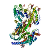
+ Open data
- Basic information
| Entry | Database: PDB / ID: 8if4 | |||||||||
|---|---|---|---|---|---|---|---|---|---|---|
| Title | Structure of human alpha-2/delta-1 without mirogabalin | |||||||||
 Components Components | Voltage-dependent calcium channel subunit alpha-2/delta-1 Voltage-gated calcium channel Voltage-gated calcium channel | |||||||||
 Keywords Keywords |  MEMBRANE PROTEIN / gabapentinoid / MEMBRANE PROTEIN / gabapentinoid /  Cache domain / Cache domain /  cryo-EM cryo-EM | |||||||||
| Function / homology |  Function and homology information Function and homology informationregulation of membrane repolarization during action potential / positive regulation of high voltage-gated calcium channel activity / calcium ion transmembrane transport via high voltage-gated calcium channel / membrane depolarization during bundle of His cell action potential /  L-type voltage-gated calcium channel complex / cardiac muscle cell action potential involved in contraction / regulation of ventricular cardiac muscle cell membrane repolarization / calcium ion transport into cytosol / regulation of calcium ion transmembrane transport via high voltage-gated calcium channel / L-type voltage-gated calcium channel complex / cardiac muscle cell action potential involved in contraction / regulation of ventricular cardiac muscle cell membrane repolarization / calcium ion transport into cytosol / regulation of calcium ion transmembrane transport via high voltage-gated calcium channel /  voltage-gated calcium channel complex ...regulation of membrane repolarization during action potential / positive regulation of high voltage-gated calcium channel activity / calcium ion transmembrane transport via high voltage-gated calcium channel / membrane depolarization during bundle of His cell action potential / voltage-gated calcium channel complex ...regulation of membrane repolarization during action potential / positive regulation of high voltage-gated calcium channel activity / calcium ion transmembrane transport via high voltage-gated calcium channel / membrane depolarization during bundle of His cell action potential /  L-type voltage-gated calcium channel complex / cardiac muscle cell action potential involved in contraction / regulation of ventricular cardiac muscle cell membrane repolarization / calcium ion transport into cytosol / regulation of calcium ion transmembrane transport via high voltage-gated calcium channel / L-type voltage-gated calcium channel complex / cardiac muscle cell action potential involved in contraction / regulation of ventricular cardiac muscle cell membrane repolarization / calcium ion transport into cytosol / regulation of calcium ion transmembrane transport via high voltage-gated calcium channel /  voltage-gated calcium channel complex / neuronal dense core vesicle / regulation of heart rate by cardiac conduction / calcium ion import across plasma membrane / regulation of calcium ion transport / voltage-gated calcium channel complex / neuronal dense core vesicle / regulation of heart rate by cardiac conduction / calcium ion import across plasma membrane / regulation of calcium ion transport /  voltage-gated calcium channel activity / voltage-gated calcium channel activity /  sarcoplasmic reticulum / cellular response to amyloid-beta / calcium ion transport / extracellular exosome / sarcoplasmic reticulum / cellular response to amyloid-beta / calcium ion transport / extracellular exosome /  metal ion binding / metal ion binding /  plasma membrane plasma membraneSimilarity search - Function | |||||||||
| Biological species |   Homo sapiens (human) Homo sapiens (human) | |||||||||
| Method |  ELECTRON MICROSCOPY / ELECTRON MICROSCOPY /  single particle reconstruction / single particle reconstruction /  cryo EM / Resolution: 3.23 Å cryo EM / Resolution: 3.23 Å | |||||||||
 Authors Authors | Kozai, D. / Numoto, N. / Fujiyoshi, Y. | |||||||||
| Funding support |  Japan, 2items Japan, 2items
| |||||||||
 Citation Citation |  Journal: J Mol Biol / Year: 2023 Journal: J Mol Biol / Year: 2023Title: Recognition Mechanism of a Novel Gabapentinoid Drug, Mirogabalin, for Recombinant Human αδ1, a Voltage-Gated Calcium Channel Subunit. Authors: Daisuke Kozai / Nobutaka Numoto / Kouki Nishikawa / Akiko Kamegawa / Shohei Kawasaki / Yoko Hiroaki / Katsumasa Irie / Atsunori Oshima / Hiroyuki Hanzawa / Kousei Shimada / Yutaka Kitano / Yoshinori Fujiyoshi /  Abstract: Mirogabalin is a novel gabapentinoid drug with a hydrophobic bicyclo substituent on the γ-aminobutyric acid moiety that targets the voltage-gated calcium channel subunit αδ1. Here, to reveal the ...Mirogabalin is a novel gabapentinoid drug with a hydrophobic bicyclo substituent on the γ-aminobutyric acid moiety that targets the voltage-gated calcium channel subunit αδ1. Here, to reveal the mirogabalin recognition mechanisms of αδ1, we present structures of recombinant human αδ1 with and without mirogabalin analyzed by cryo-electron microscopy. These structures show the binding of mirogabalin to the previously reported gabapentinoid binding site, which is the extracellular dCache_1 domain containing a conserved amino acid binding motif. A slight conformational change occurs around the residues positioned close to the hydrophobic group of mirogabalin. Mutagenesis binding assays identified that residues in the hydrophobic interaction region, in addition to several amino acid binding motif residues around the amino and carboxyl groups of mirogabalin, are critical for mirogabalin binding. The A215L mutation introduced to decrease the hydrophobic pocket volume predictably suppressed mirogabalin binding and promoted the binding of another ligand, L-Leu, with a smaller hydrophobic substituent than mirogabalin. Alterations of residues in the hydrophobic interaction region of αδ1 to those of the αδ2, αδ3, and αδ4 isoforms, of which αδ3 and αδ4 are gabapentin-insensitive, suppressed the binding of mirogabalin. These results support the importance of hydrophobic interactions in αδ1 ligand recognition. | |||||||||
| History |
|
- Structure visualization
| Structure viewer | Molecule:  Molmil Molmil Jmol/JSmol Jmol/JSmol |
|---|
- Downloads & links
- Download
| PDBx/mmCIF format |  8if4.cif.gz 8if4.cif.gz | 199.3 KB | Display |  PDBx/mmCIF format PDBx/mmCIF format |
|---|---|---|---|---|
| PDB format |  pdb8if4.ent.gz pdb8if4.ent.gz | 156.3 KB | Display |  PDB format PDB format |
| PDBx/mmJSON format |  8if4.json.gz 8if4.json.gz | Tree view |  PDBx/mmJSON format PDBx/mmJSON format | |
| Others |  Other downloads Other downloads |
-Validation report
| Arichive directory |  https://data.pdbj.org/pub/pdb/validation_reports/if/8if4 https://data.pdbj.org/pub/pdb/validation_reports/if/8if4 ftp://data.pdbj.org/pub/pdb/validation_reports/if/8if4 ftp://data.pdbj.org/pub/pdb/validation_reports/if/8if4 | HTTPS FTP |
|---|
-Related structure data
| Related structure data |  35400MC  8if3C M: map data used to model this data C: citing same article ( |
|---|---|
| Similar structure data | Similarity search - Function & homology  F&H Search F&H Search |
- Links
Links
- Assembly
| Deposited unit | 
|
|---|---|
| 1 |
|
- Components
| #1: Protein |  Voltage-gated calcium channel / Voltage-gated calcium channel subunit alpha-2/delta-1 Voltage-gated calcium channel / Voltage-gated calcium channel subunit alpha-2/delta-1Mass: 124413.062 Da / Num. of mol.: 1 Source method: isolated from a genetically manipulated source Source: (gene. exp.)   Homo sapiens (human) / Gene: CACNA2D1, CACNL2A, CCHL2A, MHS3 / Plasmid: BacMam / Cell line (production host): Expi293F / Production host: Homo sapiens (human) / Gene: CACNA2D1, CACNL2A, CCHL2A, MHS3 / Plasmid: BacMam / Cell line (production host): Expi293F / Production host:   Homo sapiens (human) / References: UniProt: P54289 Homo sapiens (human) / References: UniProt: P54289 | ||
|---|---|---|---|
| #2: Polysaccharide | 2-acetamido-2-deoxy-beta-D-glucopyranose-(1-4)-2-acetamido-2-deoxy-beta-D-glucopyranose / Mass: 424.401 Da / Num. of mol.: 1 / Mass: 424.401 Da / Num. of mol.: 1Source method: isolated from a genetically manipulated source | ||
| #3: Sugar | ChemComp-NAG /  N-Acetylglucosamine N-AcetylglucosamineHas ligand of interest | N | |
-Experimental details
-Experiment
| Experiment | Method:  ELECTRON MICROSCOPY ELECTRON MICROSCOPY |
|---|---|
| EM experiment | Aggregation state: PARTICLE / 3D reconstruction method:  single particle reconstruction single particle reconstruction |
- Sample preparation
| Component | Name: Voltage-dependent calcium channel subunit alpha-2/delta-1 Voltage-gated calcium channel Voltage-gated calcium channelType: ORGANELLE OR CELLULAR COMPONENT / Entity ID: #1 / Source: RECOMBINANT |
|---|---|
| Molecular weight | Value: 0.14 MDa / Experimental value: NO |
| Source (natural) | Organism:   Homo sapiens (human) Homo sapiens (human) |
| Source (recombinant) | Organism:   Homo sapiens (human) Homo sapiens (human) |
| Buffer solution | pH: 7.5 |
| Specimen | Embedding applied: NO / Shadowing applied: NO / Staining applied : NO / Vitrification applied : NO / Vitrification applied : YES : YES |
| Specimen support | Grid material: GOLD / Grid mesh size: 300 divisions/in. / Grid type: Quantifoil R1.2/1.3 |
Vitrification | Cryogen name: ETHANE |
- Electron microscopy imaging
| Microscopy | Model: JEOL CRYO ARM 300 |
|---|---|
| Electron gun | Electron source : :  FIELD EMISSION GUN / Accelerating voltage: 300 kV / Illumination mode: FLOOD BEAM FIELD EMISSION GUN / Accelerating voltage: 300 kV / Illumination mode: FLOOD BEAM |
| Electron lens | Mode: BRIGHT FIELD Bright-field microscopy / Nominal defocus max: 2500 nm / Nominal defocus min: 500 nm Bright-field microscopy / Nominal defocus max: 2500 nm / Nominal defocus min: 500 nm |
| Image recording | Electron dose: 68.3 e/Å2 / Detector mode: COUNTING / Film or detector model: GATAN K2 SUMMIT (4k x 4k) |
- Processing
| Software | Name: REFMAC / Version: 5.8.0352 / Classification: refinement | ||||||||||||||||||||||||||||||||||||||||||||||||||||||||||||||||||||||||||||||||||||||||||||||||||||||||||
|---|---|---|---|---|---|---|---|---|---|---|---|---|---|---|---|---|---|---|---|---|---|---|---|---|---|---|---|---|---|---|---|---|---|---|---|---|---|---|---|---|---|---|---|---|---|---|---|---|---|---|---|---|---|---|---|---|---|---|---|---|---|---|---|---|---|---|---|---|---|---|---|---|---|---|---|---|---|---|---|---|---|---|---|---|---|---|---|---|---|---|---|---|---|---|---|---|---|---|---|---|---|---|---|---|---|---|---|
| EM software |
| ||||||||||||||||||||||||||||||||||||||||||||||||||||||||||||||||||||||||||||||||||||||||||||||||||||||||||
CTF correction | Type: PHASE FLIPPING AND AMPLITUDE CORRECTION | ||||||||||||||||||||||||||||||||||||||||||||||||||||||||||||||||||||||||||||||||||||||||||||||||||||||||||
3D reconstruction | Resolution: 3.23 Å / Resolution method: FSC 0.143 CUT-OFF / Num. of particles: 331613 / Symmetry type: POINT | ||||||||||||||||||||||||||||||||||||||||||||||||||||||||||||||||||||||||||||||||||||||||||||||||||||||||||
| Refinement | Resolution: 3.2→116.28 Å / Cor.coef. Fo:Fc: 0.865 / ESU R: 0.567 Stereochemistry target values: MAXIMUM LIKELIHOOD WITH PHASES Details: HYDROGENS HAVE BEEN ADDED IN THE RIDING POSITIONS
| ||||||||||||||||||||||||||||||||||||||||||||||||||||||||||||||||||||||||||||||||||||||||||||||||||||||||||
| Solvent computation | Solvent model: PARAMETERS FOR MASK CACLULATION | ||||||||||||||||||||||||||||||||||||||||||||||||||||||||||||||||||||||||||||||||||||||||||||||||||||||||||
| Displacement parameters | Biso mean: 140.868 Å2 | ||||||||||||||||||||||||||||||||||||||||||||||||||||||||||||||||||||||||||||||||||||||||||||||||||||||||||
| Refinement step | Cycle: 1 / Total: 7424 | ||||||||||||||||||||||||||||||||||||||||||||||||||||||||||||||||||||||||||||||||||||||||||||||||||||||||||
| Refine LS restraints |
|
 Movie
Movie Controller


 PDBj
PDBj


