 Movie
Movie Controller

[English] 日本語
 Yorodumi
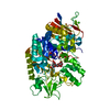
- PDB-8ids: Crystal structure of Bacillus sp. AHU2216 GH13_31 Alpha-glucosida... -
+ Open data

- Basic information
| Entry | Database: PDB / ID: 8ids | ||||||
|---|---|---|---|---|---|---|---|
| Title | Crystal structure of Bacillus sp. AHU2216 GH13_31 Alpha-glucosidase E256Q/N258P in complex with maltotriose | ||||||
 Components Components | Alpha-glucosidase | ||||||
 Keywords Keywords | HYDROLASE / alpha-glucosidase / GH13_31 / alpha-amylase / maltotriose | ||||||
| Function / homology |  Function and homology information Function and homology informationoligosaccharide catabolic process / alpha-amylase activity / metal ion bindingSimilarity search - Function | ||||||
| Biological species |  | ||||||
| Method | X-RAY DIFFRACTION / SYNCHROTRON / MOLECULAR REPLACEMENT / Resolution: 1.5 Å | ||||||
 Authors Authors | Auiewiriyanukul, W. / Saburi, W. / Yu, J. / Kato, K. / Yao, M. / Mori, H. | ||||||
| Funding support | 1items
| ||||||
 Citation Citation | Journal: Molecules / Year: 2023 Title: Alteration of Substrate Specificity and Transglucosylation Activity of GH13_31 alpha-Glucosidase from Bacillus sp. AHU2216 through Site-Directed Mutagenesis of Asn258 on beta → alpha Loop 5. Authors: Auiewiriyanukul, W. / Saburi, W. / Ota, T. / Yu, J. / Kato, K. / Yao, M. / Mori, H. | ||||||
| History |
|
- Structure visualization
| Structure viewer | Molecule: MolmilJmol/JSmol |
|---|
- Downloads & links
-Download
| PDBx/mmCIF format | 8ids.cif.gz | 141 KB | Display | PDBx/mmCIF format |
|---|---|---|---|---|
| PDB format | pdb8ids.ent.gz | 105.8 KB | Display | PDB format |
| PDBx/mmJSON format | 8ids.json.gz | Tree view | PDBx/mmJSON format | |
| Others |  Other downloads Other downloads |
-Validation report
| Arichive directory | https://data.pdbj.org/pub/pdb/validation_reports/id/8idsftp://data.pdbj.org/pub/pdb/validation_reports/id/8ids | HTTPS FTP |
|---|
-Related structure data
-Links
 PDBj
PDBj

- Assembly
| Deposited unit | 
| ||||||||
|---|---|---|---|---|---|---|---|---|---|
| 1 |
| ||||||||
| Unit cell |
|
-Components
| #1: Protein | Mass: 65982.625 Da / Num. of mol.: 1 / Mutation: E256Q,N258P Source method: isolated from a genetically manipulated source Source: (gene. exp.) Gene: BspAG13A / Production host: Escherichia coli (E. coli) / References: UniProt: A0A2Z5WH92 | ||||
|---|---|---|---|---|---|
| #2: Polysaccharide | alpha-D-glucopyranose-(1-4)-alpha-D-glucopyranose-(1-4)-alpha-D-glucopyranose  , Oligosaccharide / Class: Nutrient / Mass: 504.438 Da / Num. of mol.: 1 , Oligosaccharide / Class: Nutrient / Mass: 504.438 Da / Num. of mol.: 1Source method: isolated from a genetically manipulated source Details: oligosaccharide / References: alpha-maltotriose | ||||
| #3: Chemical |   Mass: 40.078 Da / Num. of mol.: 2 / Source method: obtained synthetically / Formula: Ca Mass: 40.078 Da / Num. of mol.: 2 / Source method: obtained synthetically / Formula: Ca#4: Water | ChemComp-HOH / | Water Mass: 18.015 Da / Num. of mol.: 545 / Source method: isolated from a natural source / Formula: H2O Mass: 18.015 Da / Num. of mol.: 545 / Source method: isolated from a natural source / Formula: H2OHas ligand of interest | Y | |
-Experimental details
-Experiment
| Experiment | Method: X-RAY DIFFRACTION / Number of used crystals: 1 |
|---|
- Sample preparation
| Crystal | Density Matthews: 2.55 Å3/Da / Density % sol: 51.84 % |
|---|---|
| Crystal grow | Temperature: 293 K / Method: vapor diffusion, hanging drop / pH: 7 Details: 10 mM maltotetraose, 0.2 M calcium chloride, and 20% polyethylene glycol 3350 |
-Data collection
| Diffraction | Mean temperature: 100 K / Serial crystal experiment: N |
|---|---|
| Diffraction source | Source: SYNCHROTRON / Site: SPring-8  / Beamline: BL41XU / Wavelength: 1 Å / Beamline: BL41XU / Wavelength: 1 Å |
| Detector | Type: DECTRIS EIGER X 16M / Detector: PIXEL / Date: Apr 25, 2018 |
| Radiation | Protocol: SINGLE WAVELENGTH / Monochromatic (M) / Laue (L): M / Scattering type: x-ray |
| Radiation wavelength | Wavelength: 1 Å / Relative weight: 1 |
| Reflection | Resolution: 1.5→50 Å / Num. obs: 104555 / % possible obs: 99.9 % / Redundancy: 13.1 % / CC1/2: 0.999 / Rrim(I) all: 0.088 / Net I/σ(I): 19.71 |
| Reflection shell | Resolution: 1.5→1.59 Å / Mean I/σ(I) obs: 2.76 / Num. unique obs: 16645 / CC1/2: 0.851 / Rrim(I) all: 0.835 / % possible all: 99.6 |
- Processing
| Software |
| |||||||||||||||||||||||||||||||||||||||||||||||||||||||||||||||||||||||||||||||||||||||||||||||||||||||||||||||||||||||||||||||||||||||||||||||||||||||||||||||||||||||||||||||||||||||||||||||||||||||||||||||||||||||||
|---|---|---|---|---|---|---|---|---|---|---|---|---|---|---|---|---|---|---|---|---|---|---|---|---|---|---|---|---|---|---|---|---|---|---|---|---|---|---|---|---|---|---|---|---|---|---|---|---|---|---|---|---|---|---|---|---|---|---|---|---|---|---|---|---|---|---|---|---|---|---|---|---|---|---|---|---|---|---|---|---|---|---|---|---|---|---|---|---|---|---|---|---|---|---|---|---|---|---|---|---|---|---|---|---|---|---|---|---|---|---|---|---|---|---|---|---|---|---|---|---|---|---|---|---|---|---|---|---|---|---|---|---|---|---|---|---|---|---|---|---|---|---|---|---|---|---|---|---|---|---|---|---|---|---|---|---|---|---|---|---|---|---|---|---|---|---|---|---|---|---|---|---|---|---|---|---|---|---|---|---|---|---|---|---|---|---|---|---|---|---|---|---|---|---|---|---|---|---|---|---|---|---|---|---|---|---|---|---|---|---|---|---|---|---|---|---|---|---|
| Refinement | Method to determine structure: MOLECULAR REPLACEMENT / Resolution: 1.5→47.74 Å / SU ML: 0.15 / Cross valid method: FREE R-VALUE / σ(F): 1.35 / Phase error: 17.06 / Stereochemistry target values: ML
| |||||||||||||||||||||||||||||||||||||||||||||||||||||||||||||||||||||||||||||||||||||||||||||||||||||||||||||||||||||||||||||||||||||||||||||||||||||||||||||||||||||||||||||||||||||||||||||||||||||||||||||||||||||||||
| Solvent computation | Shrinkage radii: 0.9 Å / VDW probe radii: 1.1 Å / Solvent model: FLAT BULK SOLVENT MODEL | |||||||||||||||||||||||||||||||||||||||||||||||||||||||||||||||||||||||||||||||||||||||||||||||||||||||||||||||||||||||||||||||||||||||||||||||||||||||||||||||||||||||||||||||||||||||||||||||||||||||||||||||||||||||||
| Refinement step | Cycle: LAST / Resolution: 1.5→47.74 Å
| |||||||||||||||||||||||||||||||||||||||||||||||||||||||||||||||||||||||||||||||||||||||||||||||||||||||||||||||||||||||||||||||||||||||||||||||||||||||||||||||||||||||||||||||||||||||||||||||||||||||||||||||||||||||||
| Refine LS restraints |
| |||||||||||||||||||||||||||||||||||||||||||||||||||||||||||||||||||||||||||||||||||||||||||||||||||||||||||||||||||||||||||||||||||||||||||||||||||||||||||||||||||||||||||||||||||||||||||||||||||||||||||||||||||||||||
| LS refinement shell |
|
