[English] 日本語
Yorodumi
- PDB-8i4r: Crystal structure of Acyl-CoA dehydrogenase complexed with Acetyl... -

+
Open data


ID or keywords:

Loading...

-
Basic information

Entry
Database: PDB / ID: 8i4r
TitleCrystal structure of Acyl-CoA dehydrogenase complexed with Acetyl-CoA from Thermobifida fusca
ComponentsAcyl-CoA dehydrogenase
KeywordsOXIDOREDUCTASE / Acyl-CoA dehydrogenase
Function / homology
Function and homology information


Oxidoreductases; Acting on the CH-CH group of donors; With a flavin as acceptor / acyl-CoA dehydrogenase activity / flavin adenine dinucleotide binding
Similarity search - Function
Acyl-CoA dehydrogenases signature 1. / Acyl-CoA dehydrogenases signature 2. / Acyl-CoA dehydrogenase, conserved site / Acyl-CoA oxidase/dehydrogenase, middle domain superfamily / Acyl-CoA dehydrogenase/oxidase C-terminal / Acyl-CoA dehydrogenase/oxidase, N-terminal / Acyl-CoA dehydrogenase, C-terminal domain / Acyl-CoA dehydrogenase, N-terminal domain / Acyl-CoA oxidase/dehydrogenase, middle domain / Acyl-CoA dehydrogenase, middle domain ...Acyl-CoA dehydrogenases signature 1. / Acyl-CoA dehydrogenases signature 2. / Acyl-CoA dehydrogenase, conserved site / Acyl-CoA oxidase/dehydrogenase, middle domain superfamily / Acyl-CoA dehydrogenase/oxidase C-terminal / Acyl-CoA dehydrogenase/oxidase, N-terminal / Acyl-CoA dehydrogenase, C-terminal domain / Acyl-CoA dehydrogenase, N-terminal domain / Acyl-CoA oxidase/dehydrogenase, middle domain / Acyl-CoA dehydrogenase, middle domain / Acyl-CoA dehydrogenase/oxidase, N-terminal domain superfamily / Acyl-CoA dehydrogenase/oxidase, N-terminal and middle domain superfamily / Acyl-CoA dehydrogenase-like, C-terminal
Similarity search - Domain/homology
ACETYL COENZYME *A / FLAVIN-ADENINE DINUCLEOTIDE / Acyl-CoA dehydrogenase
Similarity search - Component
Biological speciesThermobifida fusca (bacteria)
MethodX-RAY DIFFRACTION / SYNCHROTRON / MOLECULAR REPLACEMENT / Resolution: 2.51 Å
AuthorsChoi, M. / Seok, J. / Kim, K.-J.
Funding support1items
OrganizationGrant numberCountry
Not funded
CitationJournal: To Be Published
Title: Crystal structure of Acyl-CoA dehydrogenase complexed with Acetyl-CoA from Thermobifida fusca
Authors: Choi, M. / Kim, K.-J.
History
DepositionJan 20, 2023Deposition site: PDBJ / Processing site: PDBJ
Revision 1.0Jan 24, 2024Provider: repository / Type: Initial release

-
Structure visualization

Structure viewerMolecule:
MolmilJmol/JSmol

Downloads & links

-
Assembly

Deposited unit
A: Acyl-CoA dehydrogenase
hetero molecules


Theoretical massNumber of molelcules
Total (without water)44,9143
Polymers43,3191
Non-polymers1,5952
Water1,29772
1
A: Acyl-CoA dehydrogenase
hetero molecules

A: Acyl-CoA dehydrogenase
hetero molecules

A: Acyl-CoA dehydrogenase
hetero molecules

A: Acyl-CoA dehydrogenase
hetero molecules


Theoretical massNumber of molelcules
Total (without water)179,65612
Polymers173,2754
Non-polymers6,3808
Water724
TypeNameSymmetry operationNumber
identity operation1_555x,y,z1
crystal symmetry operation8_555-y,-x,-z1
crystal symmetry operation10_555-x,-y,z1
crystal symmetry operation15_555y,x,-z1
Buried area29290 Å2
ΔGint-159 kcal/mol
Surface area46010 Å2
MethodPISA
Unit cell
Length a, b, c (Å)102.143, 102.143, 312.183
Angle α, β, γ (deg.)90.00, 90.00, 90.00
Int Tables number98
Space group name H-MI4122
Components on special symmetry positions
IDModelComponents
11A-533-

HOH

21A-572-

HOH

-
Components

#1: Protein Acyl-CoA dehydrogenase /


Mass: 43318.863 Da / Num. of mol.: 1
Source method: isolated from a genetically manipulated source
Source: (gene. exp.) Thermobifida fusca (bacteria) / Strain: strain YX / Gene: Tfu_1647 / Plasmid: pET30a / Production host: Escherichia coli BL21(DE3) (bacteria) / References: UniProt: Q47PD7
#2: Chemical ChemComp-FAD / FLAVIN-ADENINE DINUCLEOTIDE / Flavin adenine dinucleotide


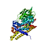
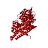
Mass: 785.550 Da / Num. of mol.: 1 / Source method: isolated from a natural source / Formula: C27H33N9O15P2 / Feature type: SUBJECT OF INVESTIGATION / Comment: FAD*YM
#3: Chemical ChemComp-ACO / ACETYL COENZYME *A / Acetyl-CoA


Mass: 809.571 Da / Num. of mol.: 1 / Source method: obtained synthetically / Formula: C23H38N7O17P3S / Feature type: SUBJECT OF INVESTIGATION
#4: Water ChemComp-HOH / water / Water


Mass: 18.015 Da / Num. of mol.: 72 / Source method: isolated from a natural source / Formula: H2O
Has ligand of interestY

-
Experimental details

-
Experiment

ExperimentMethod: X-RAY DIFFRACTION / Number of used crystals: 1

-
Sample preparation

CrystalDensity Matthews: 4.7 Å3/Da / Density % sol: 73.83 %
Crystal growTemperature: 293 K / Method: vapor diffusion, hanging drop
Details: Ammonium sulfate Sodium citrate Potassium sodium tartrate tetrahydrate

-
Data collection

DiffractionMean temperature: 100 K / Serial crystal experiment: N
Diffraction sourceSource: SYNCHROTRON / Site: PAL/PLS / Beamline: 7A (6B, 6C1) / Wavelength: 0.97934 Å
DetectorType: ADSC QUANTUM 270 / Detector: CCD / Date: May 27, 2022
RadiationProtocol: SINGLE WAVELENGTH / Monochromatic (M) / Laue (L): M / Scattering type: x-ray
Radiation wavelengthWavelength: 0.97934 Å / Relative weight: 1
ReflectionResolution: 2.5→50 Å / Num. obs: 27192 / % possible obs: 99.9 % / Redundancy: 7.4 % / Rmerge(I) obs: 0.089 / Χ2: 0.039 / Net I/σ(I): 12.8
Reflection shell
Resolution (Å)Redundancy (%)Rmerge(I) obsNum. unique obsΧ2Diffraction-ID% possible all
2.5-2.5470.31327091.9951100
2.54-2.597.10.29427132.0181100
2.59-2.647.20.27227182.091100
2.64-2.697.20.23826812.0491100
2.69-2.757.30.22127402.1151100
2.75-2.827.40.19726852.1571100
2.82-2.897.40.18527142.2151100
2.89-2.967.50.17127122.3231100
2.96-3.057.50.14327002.4121100
3.05-3.157.60.13526822.6221100
3.15-3.267.60.1227242.7611100
3.26-3.397.60.10627342.9411100
3.39-3.557.60.09726793.171100
3.55-3.737.60.08527523.3751100
3.73-3.977.60.07726813.6141100
3.97-4.277.50.07327123.7311100
4.27-4.77.50.06926993.761100
4.7-5.387.50.06627173.2111100
5.38-6.787.40.06927242.9811100
6.78-506.90.05926983.873199

-
Processing

Software
NameVersionClassification
HKL-2000data scaling
MOLREPphasing
REFMACv7.1.018refinement
MOLREPphasing
HKL-2000data reduction
RefinementMethod to determine structure: MOLECULAR REPLACEMENT / Resolution: 2.51→32.8 Å / Cor.coef. Fo:Fc: 0.96 / Cor.coef. Fo:Fc free: 0.944 / SU B: 4.951 / SU ML: 0.109 / Cross valid method: THROUGHOUT / ESU R: 0.194 / ESU R Free: 0.169 / Stereochemistry target values: MAXIMUM LIKELIHOOD / Details: HYDROGENS HAVE BEEN ADDED IN THE RIDING POSITIONS
RfactorNum. reflection% reflectionSelection details
Rfree0.19488 1426 5 %RANDOM
Rwork0.16551 ---
obs0.16701 27192 98.83 %-
Solvent computationIon probe radii: 0.8 Å / Shrinkage radii: 0.8 Å / VDW probe radii: 1.2 Å / Solvent model: MASK
Displacement parametersBiso mean: 30.744 Å2
Baniso -1Baniso -2Baniso -3
1-2.3 Å2-0 Å2-0 Å2
2--2.3 Å2-0 Å2
3----4.6 Å2
Refinement stepCycle: 1 / Resolution: 2.51→32.8 Å
ProteinNucleic acidLigandSolventTotal
Num. atoms2932 0 104 72 3108
Refine LS restraints
Refine-IDTypeDev idealDev ideal targetNumber
X-RAY DIFFRACTIONr_bond_refined_d0.0110.0123094
X-RAY DIFFRACTIONr_bond_other_d0.0010.0162790
X-RAY DIFFRACTIONr_angle_refined_deg1.8341.6494198
X-RAY DIFFRACTIONr_angle_other_deg0.6051.5566488
X-RAY DIFFRACTIONr_dihedral_angle_1_deg6.1075380
X-RAY DIFFRACTIONr_dihedral_angle_2_deg18.4319.51631
X-RAY DIFFRACTIONr_dihedral_angle_3_deg15.37610506
X-RAY DIFFRACTIONr_dihedral_angle_4_deg
X-RAY DIFFRACTIONr_chiral_restr0.0850.2460
X-RAY DIFFRACTIONr_gen_planes_refined0.0110.023564
X-RAY DIFFRACTIONr_gen_planes_other0.0010.02612
X-RAY DIFFRACTIONr_nbd_refined
X-RAY DIFFRACTIONr_nbd_other
X-RAY DIFFRACTIONr_nbtor_refined
X-RAY DIFFRACTIONr_nbtor_other
X-RAY DIFFRACTIONr_xyhbond_nbd_refined
X-RAY DIFFRACTIONr_xyhbond_nbd_other
X-RAY DIFFRACTIONr_metal_ion_refined
X-RAY DIFFRACTIONr_metal_ion_other
X-RAY DIFFRACTIONr_symmetry_vdw_refined
X-RAY DIFFRACTIONr_symmetry_vdw_other
X-RAY DIFFRACTIONr_symmetry_hbond_refined
X-RAY DIFFRACTIONr_symmetry_hbond_other
X-RAY DIFFRACTIONr_symmetry_metal_ion_refined
X-RAY DIFFRACTIONr_symmetry_metal_ion_other
X-RAY DIFFRACTIONr_mcbond_it2.6112.9151524
X-RAY DIFFRACTIONr_mcbond_other2.5922.9141523
X-RAY DIFFRACTIONr_mcangle_it3.6244.3641902
X-RAY DIFFRACTIONr_mcangle_other3.6234.3671903
X-RAY DIFFRACTIONr_scbond_it4.5023.5221570
X-RAY DIFFRACTIONr_scbond_other4.5033.5241571
X-RAY DIFFRACTIONr_scangle_it
X-RAY DIFFRACTIONr_scangle_other6.4065.1062297
X-RAY DIFFRACTIONr_long_range_B_refined7.23537.873503
X-RAY DIFFRACTIONr_long_range_B_other7.23637.8713502
X-RAY DIFFRACTIONr_rigid_bond_restr
X-RAY DIFFRACTIONr_sphericity_free
X-RAY DIFFRACTIONr_sphericity_bonded
LS refinement shellResolution: 2.51→2.571 Å
RfactorNum. reflection% reflection
Rfree0.207 101 -
Rwork0.201 1935 -
obs--97.37 %

+
About Yorodumi

-
News

-
Feb 9, 2022. New format data for meta-information of EMDB entries

New format data for meta-information of EMDB entries

  • Version 3 of the EMDB header file is now the official format.
  • The previous official version 1.9 will be removed from the archive.

Related info.:EMDB header

External links:wwPDB to switch to version 3 of the EMDB data model

-
Aug 12, 2020. Covid-19 info

Covid-19 info

URL: https://pdbj.org/emnavi/covid19.php

New page: Covid-19 featured information page in EM Navigator.

Related info.:Covid-19 info / Mar 5, 2020. Novel coronavirus structure data

+
Mar 5, 2020. Novel coronavirus structure data

Novel coronavirus structure data

Related info.:Yorodumi Speices / Aug 12, 2020. Covid-19 info

External links:COVID-19 featured content - PDBj / Molecule of the Month (242):Coronavirus Proteases

+
Jan 31, 2019. EMDB accession codes are about to change! (news from PDBe EMDB page)

EMDB accession codes are about to change! (news from PDBe EMDB page)

  • The allocation of 4 digits for EMDB accession codes will soon come to an end. Whilst these codes will remain in use, new EMDB accession codes will include an additional digit and will expand incrementally as the available range of codes is exhausted. The current 4-digit format prefixed with “EMD-” (i.e. EMD-XXXX) will advance to a 5-digit format (i.e. EMD-XXXXX), and so on. It is currently estimated that the 4-digit codes will be depleted around Spring 2019, at which point the 5-digit format will come into force.
  • The EM Navigator/Yorodumi systems omit the EMD- prefix.

Related info.:Q: What is EMD? / ID/Accession-code notation in Yorodumi/EM Navigator

External links:EMDB Accession Codes are Changing Soon! / Contact to PDBj

+
Jul 12, 2017. Major update of PDB

Major update of PDB

  • wwPDB released updated PDB data conforming to the new PDBx/mmCIF dictionary.
  • This is a major update changing the version number from 4 to 5, and with Remediation, in which all the entries are updated.
  • In this update, many items about electron microscopy experimental information are reorganized (e.g. em_software).
  • Now, EM Navigator and Yorodumi are based on the updated data.

External links:wwPDB Remediation / Enriched Model Files Conforming to OneDep Data Standards Now Available in the PDB FTP Archive

-
Yorodumi

Thousand views of thousand structures

  • Yorodumi is a browser for structure data from EMDB, PDB, SASBDB, etc.
  • This page is also the successor to EM Navigator detail page, and also detail information page/front-end page for Omokage search.
  • The word "yorodu" (or yorozu) is an old Japanese word meaning "ten thousand". "mi" (miru) is to see.

Related info.:EMDB / PDB / SASBDB / Comparison of 3 databanks / Yorodumi Search / Aug 31, 2016. New EM Navigator & Yorodumi / Yorodumi Papers / Jmol/JSmol / Function and homology information / Changes in new EM Navigator and Yorodumi

Read more