 Movie
Movie Controller

[English] 日本語
 Yorodumi
- PDB-8i0q: Structure of beta-arrestin1 in complex with a phosphopeptide corr... -
+ Open data

- Basic information
| Entry | Database: PDB / ID: 8i0q | ||||||
|---|---|---|---|---|---|---|---|
| Title | Structure of beta-arrestin1 in complex with a phosphopeptide corresponding to the human C-X-C chemokine receptor type 4, CXCR4 (Local refine) | ||||||
 Components Components |
| ||||||
 Keywords Keywords |  SIGNALING PROTEIN/IMMUNE SYSTEM / GPCR / Arrestin / SIGNALING PROTEIN / SIGNALING PROTEIN-IMMUNE SYSTEM complex SIGNALING PROTEIN/IMMUNE SYSTEM / GPCR / Arrestin / SIGNALING PROTEIN / SIGNALING PROTEIN-IMMUNE SYSTEM complex | ||||||
| Function / homology |  Function and homology informationV2 vasopressin receptor binding / alpha-1A adrenergic receptor binding / follicle-stimulating hormone receptor binding / Activation of SMO / sensory perception of touch / C-X-C motif chemokine 12 receptor activity / regulation of viral process / G alpha (s) signalling events / alpha-1B adrenergic receptor binding / positive regulation of vascular wound healing ...V2 vasopressin receptor binding / alpha-1A adrenergic receptor binding / follicle-stimulating hormone receptor binding / Activation of SMO / sensory perception of touch / C-X-C motif chemokine 12 receptor activity / regulation of viral process / G alpha (s) signalling events / alpha-1B adrenergic receptor binding / positive regulation of vascular wound healing / positive regulation of macrophage migration inhibitory factor signaling pathway / follicle-stimulating hormone signaling pathway / protein phosphorylated amino acid binding / positive regulation of mesenchymal stem cell migration / angiotensin receptor binding / neuron recognition / response to ultrasound / response to tacrolimus / telencephalon cell migration / C-X-C chemokine receptor activity / Specification of primordial germ cells / CXCL12-activated CXCR4 signaling pathway / Lysosome Vesicle Biogenesis / AP-2 adaptor complex binding / myosin light chain binding / Golgi Associated Vesicle Biogenesis / MAP2K and MAPK activation / Ub-specific processing proteases / myelin maintenance / positive regulation of vasculature development / regulation of programmed cell death / positive regulation of smooth muscle cell apoptotic process / C-C chemokine receptor activity / endothelial tube morphogenesis / endothelial cell differentiation / negative regulation of interleukin-8 production / Cargo recognition for clathrin-mediated endocytosis / C-C chemokine binding / Signaling by ROBO receptors / Clathrin-mediated endocytosis / clathrin adaptor activity / regulation of chemotaxis / positive regulation of chemotaxis / cellular response to organonitrogen compound / Formation of definitive endoderm / positive regulation of dendrite extension / regulation of G protein-coupled receptor signaling pathway / arrestin family protein binding / G protein-coupled receptor internalization / anchoring junction / Thrombin signalling through proteinase activated receptors (PARs) / Chemokine receptors bind chemokines / dendritic cell chemotaxis / mitogen-activated protein kinase kinase binding / positive regulation of Rho protein signal transduction / clathrin binding / positive regulation of oligodendrocyte differentiation / stress fiber assembly / negative regulation of Notch signaling pathway / epithelial cell development / cell leading edge / pseudopodium / cellular response to cytokine stimulus / positive regulation of insulin secretion involved in cellular response to glucose stimulus / cysteine-type endopeptidase inhibitor activity involved in apoptotic process / detection of temperature stimulus involved in sensory perception of pain / small molecule binding / negative regulation of interleukin-6 production / regulation of calcium ion transport / positive regulation of receptor internalization / phototransduction / Binding and entry of HIV virion / coreceptor activity / detection of mechanical stimulus involved in sensory perception of pain / regulation of cell adhesion / clathrin-coated pit / negative regulation of protein ubiquitination / cardiac muscle contraction / insulin-like growth factor receptor binding / visual perception / neurogenesis / GTPase activator activity / cell chemotaxis / negative regulation of protein phosphorylation / response to activity / ubiquitin binding / positive regulation of protein ubiquitination / G protein-coupled receptor binding / G protein-coupled receptor activity / nuclear estrogen receptor binding / calcium-mediated signaling / phosphoprotein binding / brain development / neuron migration / response to virus / adenylate cyclase-modulating G protein-coupled receptor signaling pathway / negative regulation of ERK1 and ERK2 cascade / endocytosis / cellular response to xenobiotic stimulus / late endosome Function and homology informationV2 vasopressin receptor binding / alpha-1A adrenergic receptor binding / follicle-stimulating hormone receptor binding / Activation of SMO / sensory perception of touch / C-X-C motif chemokine 12 receptor activity / regulation of viral process / G alpha (s) signalling events / alpha-1B adrenergic receptor binding / positive regulation of vascular wound healing ...V2 vasopressin receptor binding / alpha-1A adrenergic receptor binding / follicle-stimulating hormone receptor binding / Activation of SMO / sensory perception of touch / C-X-C motif chemokine 12 receptor activity / regulation of viral process / G alpha (s) signalling events / alpha-1B adrenergic receptor binding / positive regulation of vascular wound healing / positive regulation of macrophage migration inhibitory factor signaling pathway / follicle-stimulating hormone signaling pathway / protein phosphorylated amino acid binding / positive regulation of mesenchymal stem cell migration / angiotensin receptor binding / neuron recognition / response to ultrasound / response to tacrolimus / telencephalon cell migration / C-X-C chemokine receptor activity / Specification of primordial germ cells / CXCL12-activated CXCR4 signaling pathway / Lysosome Vesicle Biogenesis / AP-2 adaptor complex binding / myosin light chain binding / Golgi Associated Vesicle Biogenesis / MAP2K and MAPK activation / Ub-specific processing proteases / myelin maintenance / positive regulation of vasculature development / regulation of programmed cell death / positive regulation of smooth muscle cell apoptotic process / C-C chemokine receptor activity / endothelial tube morphogenesis / endothelial cell differentiation / negative regulation of interleukin-8 production / Cargo recognition for clathrin-mediated endocytosis / C-C chemokine binding / Signaling by ROBO receptors / Clathrin-mediated endocytosis / clathrin adaptor activity / regulation of chemotaxis / positive regulation of chemotaxis / cellular response to organonitrogen compound / Formation of definitive endoderm / positive regulation of dendrite extension / regulation of G protein-coupled receptor signaling pathway / arrestin family protein binding / G protein-coupled receptor internalization / anchoring junction / Thrombin signalling through proteinase activated receptors (PARs) / Chemokine receptors bind chemokines / dendritic cell chemotaxis / mitogen-activated protein kinase kinase binding / positive regulation of Rho protein signal transduction / clathrin binding / positive regulation of oligodendrocyte differentiation / stress fiber assembly / negative regulation of Notch signaling pathway / epithelial cell development / cell leading edge / pseudopodium / cellular response to cytokine stimulus / positive regulation of insulin secretion involved in cellular response to glucose stimulus / cysteine-type endopeptidase inhibitor activity involved in apoptotic process / detection of temperature stimulus involved in sensory perception of pain / small molecule binding / negative regulation of interleukin-6 production / regulation of calcium ion transport / positive regulation of receptor internalization / phototransduction / Binding and entry of HIV virion / coreceptor activity / detection of mechanical stimulus involved in sensory perception of pain / regulation of cell adhesion / clathrin-coated pit / negative regulation of protein ubiquitination / cardiac muscle contraction / insulin-like growth factor receptor binding / visual perception / neurogenesis / GTPase activator activity / cell chemotaxis / negative regulation of protein phosphorylation / response to activity / ubiquitin binding / positive regulation of protein ubiquitination / G protein-coupled receptor binding / G protein-coupled receptor activity / nuclear estrogen receptor binding / calcium-mediated signaling / phosphoprotein binding / brain development / neuron migration / response to virus / adenylate cyclase-modulating G protein-coupled receptor signaling pathway / negative regulation of ERK1 and ERK2 cascade / endocytosis / cellular response to xenobiotic stimulus / late endosomeSimilarity search - Function | ||||||
| Biological species |  Rattus norvegicus (Norway rat)Mus musculus (house mouse) Rattus norvegicus (Norway rat)Mus musculus (house mouse) Homo sapiens (human) Homo sapiens (human) | ||||||
| Method | ELECTRON MICROSCOPY / single particle reconstruction / cryo EM / Resolution: 4.45 Å | ||||||
 Authors Authors | Maharana, J. / Sarma, P. / Yadav, M.K. / Banerjee, R. / Shukla, A.K. | ||||||
| Funding support |  India, 1items India, 1items
| ||||||
 Citation Citation | Journal: Mol.Cell / Year: 2023 Title: Structure of beta-arrestin in complex with a phosphopeptide Authors: Maharana, J. / Sarma, P. / Yadav, M.K. / Banerjee, R. / Shukla, A.K. | ||||||
| History |
|
- Structure visualization
| Structure viewer | Molecule: MolmilJmol/JSmol |
|---|
- Downloads & links
-Download
| PDBx/mmCIF format | 8i0q.cif.gz | 212.6 KB | Display | PDBx/mmCIF format |
|---|---|---|---|---|
| PDB format | pdb8i0q.ent.gz | 167.6 KB | Display | PDB format |
| PDBx/mmJSON format | 8i0q.json.gz | Tree view | PDBx/mmJSON format | |
| Others |  Other downloads Other downloads |
-Validation report
| Arichive directory | https://data.pdbj.org/pub/pdb/validation_reports/i0/8i0qftp://data.pdbj.org/pub/pdb/validation_reports/i0/8i0q | HTTPS FTP |
|---|
-Related structure data
| Related structure data |  35106MC  8go8C  8gocC 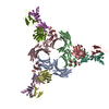 8gooC 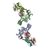 8gp3C 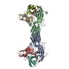 8i0nC 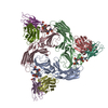 8i0zC 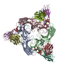 8i10C M: map data used to model this data C: citing same article ( |
|---|---|
| Similar structure data |


-Links
 PDBj
PDBj


- Assembly
| Deposited unit | 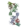
|
|---|---|
| 1 |
|
-Components
| #1: Protein | Arrestin / Arrestin beta-1 Mass: 47088.508 Da / Num. of mol.: 2 Source method: isolated from a genetically manipulated source Source: (gene. exp.) Rattus norvegicus (Norway rat) / Gene: Arrb1 / Production host:  Escherichia coli (E. coli) / References: UniProt: P29066 Escherichia coli (E. coli) / References: UniProt: P29066#2: Antibody | Mass: 25512.354 Da / Num. of mol.: 2 Source method: isolated from a genetically manipulated source Source: (gene. exp.) Mus musculus (house mouse) / Production host: Escherichia coli (E. coli)#3: Antibody | Mass: 23435.064 Da / Num. of mol.: 2 Source method: isolated from a genetically manipulated source Source: (gene. exp.) Mus musculus (house mouse) / Production host: Escherichia coli (E. coli)#4: Protein/peptide | Mass: 2380.530 Da / Num. of mol.: 2 / Source method: obtained synthetically / Source: (synth.) Homo sapiens (human) / References: UniProt: P61073Has ligand of interest | Y | |
|---|
-Experimental details
-Experiment
| Experiment | Method: ELECTRON MICROSCOPY |
|---|---|
| EM experiment | Aggregation state: PARTICLE / 3D reconstruction method: single particle reconstruction |
- Sample preparation
| Component |
| |||||||||||||||||||||||||||||||||||
|---|---|---|---|---|---|---|---|---|---|---|---|---|---|---|---|---|---|---|---|---|---|---|---|---|---|---|---|---|---|---|---|---|---|---|---|---|
| Molecular weight | Value: 0.19 MDa / Experimental value: YES | |||||||||||||||||||||||||||||||||||
| Source (natural) |
| |||||||||||||||||||||||||||||||||||
| Source (recombinant) |
| |||||||||||||||||||||||||||||||||||
| Buffer solution | pH: 7.4 | |||||||||||||||||||||||||||||||||||
| Buffer component |
| |||||||||||||||||||||||||||||||||||
| Specimen | Embedding applied: NO / Shadowing applied: NO / Staining applied: NO / Vitrification applied: YES | |||||||||||||||||||||||||||||||||||
| Vitrification | Instrument: LEICA EM GP / Cryogen name: ETHANE / Humidity: 90 % / Chamber temperature: 283.15 K / Details: Blotted for 3 seconds before plunging. |
- Electron microscopy imaging
| Microscopy | Model: TFS GLACIOS |
|---|---|
| Electron gun | Electron source: FIELD EMISSION GUN / Accelerating voltage: 200 kV / Illumination mode: FLOOD BEAM |
| Electron lens | Mode: BRIGHT FIELDBright-field microscopy / Nominal magnification: 46000 X / Nominal defocus max: 2500 nm / Nominal defocus min: 500 nm / Cs: 2.7 mm / Alignment procedure: COMA FREE |
| Specimen holder | Cryogen: NITROGEN |
| Image recording | Electron dose: 49.3 e/Å2 / Detector mode: COUNTING / Film or detector model: GATAN K3 (6k x 4k) / Num. of real images: 5637 |
| Image scans | Movie frames/image: 40 |
- Processing
| EM software |
| ||||||||||||||||||||||||||||
|---|---|---|---|---|---|---|---|---|---|---|---|---|---|---|---|---|---|---|---|---|---|---|---|---|---|---|---|---|---|
| CTF correction | Type: NONE | ||||||||||||||||||||||||||||
| Particle selection | Num. of particles selected: 3236193 | ||||||||||||||||||||||||||||
| Symmetry | Point symmetry: C2 (2 fold cyclic) | ||||||||||||||||||||||||||||
| 3D reconstruction | Resolution: 4.45 Å / Resolution method: FSC 0.143 CUT-OFF / Num. of particles: 86525 / Symmetry type: POINT | ||||||||||||||||||||||||||||
| Atomic model building | Protocol: FLEXIBLE FIT / Space: REAL | ||||||||||||||||||||||||||||
| Atomic model building | PDB-ID: 8GP3 |
