 Movie
Movie Controller

[English] 日本語
 Yorodumi
- PDB-8hv3: Crystal structure of EGFR_DMX in complex with covalently bound fr... -
+ Open data

- Basic information
| Entry | Database: PDB / ID: 8hv3 | ||||||
|---|---|---|---|---|---|---|---|
| Title | Crystal structure of EGFR_DMX in complex with covalently bound fragment 4 | ||||||
 Components Components | Epidermal growth factor receptor | ||||||
 Keywords Keywords | TRANSFERASE / Epidermal growth factor receptor kinase / covalent fragments / mutant EGFR / anti-cancer | ||||||
| Function / homology |  Function and homology information Function and homology informationresponse to hydroxyisoflavone / multivesicular body, internal vesicle lumen / positive regulation of prolactin secretion / negative regulation of cardiocyte differentiation / positive regulation of protein kinase C activity / diterpenoid metabolic process / Shc-EGFR complex / ovulation cycle / Inhibition of Signaling by Overexpressed EGFR / epidermal growth factor receptor activity ...response to hydroxyisoflavone / multivesicular body, internal vesicle lumen / positive regulation of prolactin secretion / negative regulation of cardiocyte differentiation / positive regulation of protein kinase C activity / diterpenoid metabolic process / Shc-EGFR complex / ovulation cycle / Inhibition of Signaling by Overexpressed EGFR / epidermal growth factor receptor activity / EGFR interacts with phospholipase C-gamma / positive regulation of mucus secretion / response to UV-A / epidermal growth factor binding / PLCG1 events in ERBB2 signaling / tongue development / midgut development / ERBB2-EGFR signaling pathway / hydrogen peroxide metabolic process / PTK6 promotes HIF1A stabilization / digestive tract morphogenesis / regulation of nitric-oxide synthase activity / ERBB2 Activates PTK6 Signaling / morphogenesis of an epithelial fold / intracellular vesicle / Signaling by EGFR / response to cobalamin / negative regulation of epidermal growth factor receptor signaling pathway / transmembrane receptor protein tyrosine kinase activator activity / protein tyrosine kinase activator activity / regulation of phosphatidylinositol 3-kinase/protein kinase B signal transduction / Signaling by ERBB4 / eyelid development in camera-type eye / protein insertion into membrane / cerebral cortex cell migration / ERBB2 Regulates Cell Motility / regulation of JNK cascade / : / PI3K events in ERBB2 signaling / positive regulation of cyclin-dependent protein serine/threonine kinase activity / negative regulation of mitotic cell cycle / hair follicle development / MAP kinase kinase kinase activity / Estrogen-dependent nuclear events downstream of ESR-membrane signaling / embryonic placenta development / positive regulation of bone resorption / positive regulation of G1/S transition of mitotic cell cycle / GAB1 signalosome / positive regulation of nitric oxide mediated signal transduction / salivary gland morphogenesis / peptidyl-tyrosine autophosphorylation / regulation of peptidyl-tyrosine phosphorylation / positive regulation of glial cell proliferation / positive regulation of phosphorylation / positive regulation of vasoconstriction / Signaling by ERBB2 / cellular response to epidermal growth factor stimulus / cellular response to cadmium ion / GRB2 events in EGFR signaling / SHC1 events in EGFR signaling / EGFR Transactivation by Gastrin / positive regulation of DNA repair / TFAP2 (AP-2) family regulates transcription of growth factors and their receptors / GRB2 events in ERBB2 signaling / transmembrane receptor protein tyrosine kinase activity / neurogenesis / SHC1 events in ERBB2 signaling / cellular response to dexamethasone stimulus / ossification / positive regulation of synaptic transmission, glutamatergic / regulation of ERK1 and ERK2 cascade / basal plasma membrane / neuron projection morphogenesis / positive regulation of superoxide anion generation / epithelial cell proliferation / positive regulation of DNA replication / Signal transduction by L1 / cellular response to estradiol stimulus / positive regulation of epithelial cell proliferation / NOTCH3 Activation and Transmission of Signal to the Nucleus / liver regeneration / astrocyte activation / cellular response to amino acid stimulus / positive regulation of protein localization to plasma membrane / EGFR downregulation / positive regulation of smooth muscle cell proliferation / lung development / Signaling by ERBB2 TMD/JMD mutants / clathrin-coated endocytic vesicle membrane / Constitutive Signaling by EGFRvIII / positive regulation of MAP kinase activity / Signaling by ERBB2 ECD mutants / epidermal growth factor receptor signaling pathway / Signaling by ERBB2 KD Mutants / negative regulation of protein catabolic process / receptor protein-tyrosine kinase / cell-cell adhesion / Downregulation of ERBB2 signaling / ruffle membrane / cellular response to reactive oxygen speciesSimilarity search - Function | ||||||
| Biological species |  Homo sapiens (human) Homo sapiens (human) | ||||||
| Method | X-RAY DIFFRACTION / MOLECULAR REPLACEMENT / Resolution: 2.4 Å | ||||||
 Authors Authors | Dokurno, P. | ||||||
| Funding support | 1items
| ||||||
 Citation Citation | Journal: Rsc Med Chem / Year: 2023 Title: A covalent fragment-based strategy targeting a novel cysteine to inhibit activity of mutant EGFR kinase. Authors: Kuki, N. / Walmsley, D.L. / Kanai, K. / Takechi, S. / Yoshida, M. / Murakami, R. / Takano, K. / Tominaga, Y. / Takahashi, M. / Ito, S. / Nakao, N. / Angove, H. / Baker, L.M. / Carter, E. / ...Authors: Kuki, N. / Walmsley, D.L. / Kanai, K. / Takechi, S. / Yoshida, M. / Murakami, R. / Takano, K. / Tominaga, Y. / Takahashi, M. / Ito, S. / Nakao, N. / Angove, H. / Baker, L.M. / Carter, E. / Dokurno, P. / Le Strat, L. / Macias, A.T. / Molyneaux, C.A. / Murray, J.B. / Surgenor, A.E. / Hamada, T. / Hubbard, R.E. | ||||||
| History |
|
- Structure visualization
| Structure viewer | Molecule: MolmilJmol/JSmol |
|---|
- Downloads & links
-Download
| PDBx/mmCIF format | 8hv3.cif.gz | 82.4 KB | Display | PDBx/mmCIF format |
|---|---|---|---|---|
| PDB format | pdb8hv3.ent.gz | 58.7 KB | Display | PDB format |
| PDBx/mmJSON format | 8hv3.json.gz | Tree view | PDBx/mmJSON format | |
| Others |  Other downloads Other downloads |
-Validation report
| Arichive directory | https://data.pdbj.org/pub/pdb/validation_reports/hv/8hv3ftp://data.pdbj.org/pub/pdb/validation_reports/hv/8hv3 | HTTPS FTP |
|---|
-Related structure data
| Related structure data |  8hv1C  8hv2C  8hv4C  8hv5C 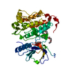 8hv6C 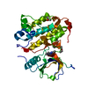 8hv7C 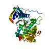 8hv8C 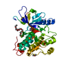 8hv9C 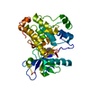 8hvaC 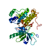 5edqS S: Starting model for refinement C: citing same article ( |
|---|---|
| Similar structure data |
-Links
 PDBj
PDBj


- Assembly
| Deposited unit | 
| ||||||||
|---|---|---|---|---|---|---|---|---|---|
| 1 |
| ||||||||
| Unit cell |
| ||||||||
| Components on special symmetry positions |
|
-Components
| #1: Protein | / Proto-oncogene c-ErbB-1 / Receptor tyrosine-protein kinase erbB-1 Mass: 37549.398 Da / Num. of mol.: 1 / Mutation: T790M, L858R, E865A, E866A, K867A Source method: isolated from a genetically manipulated source Source: (gene. exp.) Homo sapiens (human) / Gene: EGFR, ERBB, ERBB1, HER1 / Production host:   Spodoptera frugiperda (fall armyworm) Spodoptera frugiperda (fall armyworm)References: UniProt: P00533, receptor protein-tyrosine kinase |
|---|---|
| #2: Chemical | ChemComp-PE5 / Polyethylene glycol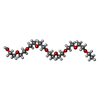  Mass: 398.489 Da / Num. of mol.: 1 / Source method: obtained synthetically / Formula: C18H38O9 / Comment: precipitant*YM Mass: 398.489 Da / Num. of mol.: 1 / Source method: obtained synthetically / Formula: C18H38O9 / Comment: precipitant*YM |
| #3: Chemical | ChemComp-DMS / Dimethyl sulfoxide  Mass: 78.133 Da / Num. of mol.: 1 / Source method: obtained synthetically / Formula: C2H6OS / Comment: DMSO, precipitant*YM Mass: 78.133 Da / Num. of mol.: 1 / Source method: obtained synthetically / Formula: C2H6OS / Comment: DMSO, precipitant*YM |
| #4: Chemical | ChemComp-MWU / ~{  Mass: 148.162 Da / Num. of mol.: 1 / Source method: obtained synthetically / Formula: C8H8N2O / Feature type: SUBJECT OF INVESTIGATION Mass: 148.162 Da / Num. of mol.: 1 / Source method: obtained synthetically / Formula: C8H8N2O / Feature type: SUBJECT OF INVESTIGATION |
| #5: Water | ChemComp-HOH / Water Mass: 18.015 Da / Num. of mol.: 134 / Source method: isolated from a natural source / Formula: H2O Mass: 18.015 Da / Num. of mol.: 134 / Source method: isolated from a natural source / Formula: H2O |
| Has ligand of interest | Y |
-Experimental details
-Experiment
| Experiment | Method: X-RAY DIFFRACTION / Number of used crystals: 1 |
|---|
- Sample preparation
| Crystal | Density Matthews: 3.39 Å3/Da / Density % sol: 63.67 % |
|---|---|
| Crystal grow | Temperature: 292 K / Method: vapor diffusion, hanging drop / pH: 8.5 / Details: 0.1M TRIS pH 8.5, 0.15M Sodium citrate, 25%PEG400 |
-Data collection
| Diffraction | Mean temperature: 110 K / Serial crystal experiment: N |
|---|---|
| Diffraction source | Source: ROTATING ANODE / Type: BRUKER TURBO X-RAY SOURCE / Wavelength: 1.54184 Å |
| Detector | Type: BRUKER PHOTON 100 / Detector: CMOS / Date: Jan 24, 2019 |
| Radiation | Protocol: SINGLE WAVELENGTH / Monochromatic (M) / Laue (L): M / Scattering type: x-ray |
| Radiation wavelength | Wavelength: 1.54184 Å / Relative weight: 1 |
| Reflection | Resolution: 2.3→21.87 Å / Num. obs: 22648 / % possible obs: 99.8 % / Redundancy: 7.05 % / Rmerge(I) obs: 0.1621 / Net I/σ(I): 9.3 |
| Reflection shell | Resolution: 2.3→2.4 Å / Redundancy: 4.6 % / Rmerge(I) obs: 0.8204 / Mean I/σ(I) obs: 1.35 / Num. unique obs: 2698 / % possible all: 100 |
- Processing
| Software |
| ||||||||||||||||||||||||||||||||||||||||||||||||||||||||||||||||||||||||||||||||||||||||||||||||||||||||||||||||||||||||||||||||||||||||||||||||||||||||||||||||||||||||||||||||||||||
|---|---|---|---|---|---|---|---|---|---|---|---|---|---|---|---|---|---|---|---|---|---|---|---|---|---|---|---|---|---|---|---|---|---|---|---|---|---|---|---|---|---|---|---|---|---|---|---|---|---|---|---|---|---|---|---|---|---|---|---|---|---|---|---|---|---|---|---|---|---|---|---|---|---|---|---|---|---|---|---|---|---|---|---|---|---|---|---|---|---|---|---|---|---|---|---|---|---|---|---|---|---|---|---|---|---|---|---|---|---|---|---|---|---|---|---|---|---|---|---|---|---|---|---|---|---|---|---|---|---|---|---|---|---|---|---|---|---|---|---|---|---|---|---|---|---|---|---|---|---|---|---|---|---|---|---|---|---|---|---|---|---|---|---|---|---|---|---|---|---|---|---|---|---|---|---|---|---|---|---|---|---|---|---|
| Refinement | Method to determine structure: MOLECULAR REPLACEMENT Starting model: 5EDQ Resolution: 2.4→20 Å / Cor.coef. Fo:Fc: 0.944 / Cor.coef. Fo:Fc free: 0.932 / SU B: 8.193 / SU ML: 0.183 / Cross valid method: THROUGHOUT / ESU R: 0.282 / ESU R Free: 0.225 / Stereochemistry target values: MAXIMUM LIKELIHOOD / Details: HYDROGENS HAVE BEEN ADDED IN THE RIDING POSITIONS
| ||||||||||||||||||||||||||||||||||||||||||||||||||||||||||||||||||||||||||||||||||||||||||||||||||||||||||||||||||||||||||||||||||||||||||||||||||||||||||||||||||||||||||||||||||||||
| Solvent computation | Ion probe radii: 0.8 Å / Shrinkage radii: 0.8 Å / VDW probe radii: 1.2 Å / Solvent model: MASK | ||||||||||||||||||||||||||||||||||||||||||||||||||||||||||||||||||||||||||||||||||||||||||||||||||||||||||||||||||||||||||||||||||||||||||||||||||||||||||||||||||||||||||||||||||||||
| Displacement parameters | Biso mean: 44.518 Å2
| ||||||||||||||||||||||||||||||||||||||||||||||||||||||||||||||||||||||||||||||||||||||||||||||||||||||||||||||||||||||||||||||||||||||||||||||||||||||||||||||||||||||||||||||||||||||
| Refinement step | Cycle: 1 / Resolution: 2.4→20 Å
| ||||||||||||||||||||||||||||||||||||||||||||||||||||||||||||||||||||||||||||||||||||||||||||||||||||||||||||||||||||||||||||||||||||||||||||||||||||||||||||||||||||||||||||||||||||||
| Refine LS restraints |
|