 Movie
Movie Controller

[English] 日本語
 Yorodumi
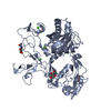
- PDB-8hq2: Crystal structure of human ADAM22 in complex with human LGI1 mutant -
+ Open data

- Basic information
| Entry | Database: PDB / ID: 8hq2 | ||||||
|---|---|---|---|---|---|---|---|
| Title | Crystal structure of human ADAM22 in complex with human LGI1 mutant | ||||||
 Components Components |
| ||||||
 Keywords Keywords |  MEMBRANE PROTEIN / SYNAPTIC MODULATOR / COMPLEX MEMBRANE PROTEIN / SYNAPTIC MODULATOR / COMPLEX | ||||||
| Function / homology |  Function and homology information Function and homology informationLGI-ADAM interactions / axon initial segment / negative regulation of cell adhesion / neurotransmitter receptor localization to postsynaptic specialization membrane / positive regulation of synaptic transmission / synaptic cleft / myelination / central nervous system development / axon guidance / metalloendopeptidase activity ...LGI-ADAM interactions / axon initial segment / negative regulation of cell adhesion / neurotransmitter receptor localization to postsynaptic specialization membrane / positive regulation of synaptic transmission / synaptic cleft / myelination / central nervous system development / axon guidance / metalloendopeptidase activity / neuron projection development / integrin binding / nervous system development / positive regulation of cell growth / cell adhesion / axon / signaling receptor binding / glutamatergic synapse / dendrite / Golgi apparatus / endoplasmic reticulum / proteolysis / extracellular space / extracellular region / membrane / plasma membrane / cytoplasmSimilarity search - Function | ||||||
| Biological species |  Homo sapiens (human) Homo sapiens (human) | ||||||
| Method | X-RAY DIFFRACTION / SYNCHROTRON / MOLECULAR REPLACEMENT / Resolution: 2.93 Å | ||||||
 Authors Authors | Liu, H. / Lin, Z. / Xu, F. | ||||||
| Funding support |  China, 1items China, 1items
| ||||||
 Citation Citation | Journal: To Be Published Title: Crystal structure of human ADAM22 in complex with human LGI1 mutant Authors: Liu, H. / Lin, Z. / Xu, F. | ||||||
| History |
|
- Structure visualization
| Structure viewer | Molecule: MolmilJmol/JSmol |
|---|
- Downloads & links
-Download
| PDBx/mmCIF format | 8hq2.cif.gz | 825 KB | Display | PDBx/mmCIF format |
|---|---|---|---|---|
| PDB format | pdb8hq2.ent.gz | 683.4 KB | Display | PDB format |
| PDBx/mmJSON format | 8hq2.json.gz | Tree view | PDBx/mmJSON format | |
| Others |  Other downloads Other downloads |
-Validation report
| Arichive directory | https://data.pdbj.org/pub/pdb/validation_reports/hq/8hq2ftp://data.pdbj.org/pub/pdb/validation_reports/hq/8hq2 | HTTPS FTP |
|---|
-Related structure data


-Links
 PDBj
PDBj


- Assembly
| Deposited unit | 
| ||||||||
|---|---|---|---|---|---|---|---|---|---|
| 1 | 
| ||||||||
| 2 | 
| ||||||||
| Unit cell |
|
-Components
| #1: Protein | Mass: 53743.102 Da / Num. of mol.: 2 Source method: isolated from a genetically manipulated source Source: (gene. exp.) Homo sapiens (human) / Gene: ADAM22, MDC2 / Production host: Homo sapiens (human) / References: UniProt: Q9P0K1#2: Protein | Mass: 60512.379 Da / Num. of mol.: 2 / Mutation: L88N/F89A/R474Q Source method: isolated from a genetically manipulated source Source: (gene. exp.) Homo sapiens (human) / Gene: LGI1, EPT, UNQ775/PRO1569 / Production host: Homo sapiens (human) / References: UniProt: O95970#3: Chemical | ChemComp-CA /   Mass: 40.078 Da / Num. of mol.: 8 / Source method: obtained synthetically / Formula: Ca Mass: 40.078 Da / Num. of mol.: 8 / Source method: obtained synthetically / Formula: Ca#4: Sugar | ChemComp-NAG / N-Acetylglucosamine  Type: D-saccharide, beta linking / Mass: 221.208 Da / Num. of mol.: 12 Type: D-saccharide, beta linking / Mass: 221.208 Da / Num. of mol.: 12Source method: isolated from a genetically manipulated source Formula: C8H15NO6 #5: Water | ChemComp-HOH / | Water Mass: 18.015 Da / Num. of mol.: 174 / Source method: isolated from a natural source / Formula: H2O Mass: 18.015 Da / Num. of mol.: 174 / Source method: isolated from a natural source / Formula: H2OHas ligand of interest | N | |
|---|
-Experimental details
-Experiment
| Experiment | Method: X-RAY DIFFRACTION / Number of used crystals: 1 |
|---|
- Sample preparation
| Crystal | Density Matthews: 2.95 Å3/Da / Density % sol: 58.35 % |
|---|---|
| Crystal grow | Temperature: 293 K / Method: vapor diffusion, sitting drop / pH: 7.5 Details: 7.5% PEG 4000, 0.2M MAGNESIUM CHLORIDE, 0.1M HEPES PH=7.5 |
-Data collection
| Diffraction | Mean temperature: 100 K / Serial crystal experiment: N |
|---|---|
| Diffraction source | Source: SYNCHROTRON / Site: SSRF / Beamline: BL19U1 / Wavelength: 0.9785 Å |
| Detector | Type: DECTRIS PILATUS3 S 6M / Detector: PIXEL / Date: May 6, 2016 |
| Radiation | Protocol: SINGLE WAVELENGTH / Monochromatic (M) / Laue (L): M / Scattering type: x-ray |
| Radiation wavelength | Wavelength: 0.9785 Å / Relative weight: 1 |
| Reflection | Resolution: 2.93→50 Å / Num. obs: 54802 / % possible obs: 98.4 % / Redundancy: 6.3 % / Rmerge(I) obs: 0.094 / Rsym value: 0.064 / Net I/σ(I): 21.167 |
| Reflection shell | Resolution: 2.93→2.99 Å / Redundancy: 6.2 % / Rmerge(I) obs: 0.944 / Mean I/σ(I) obs: 2.125 / Num. unique obs: 2716 / Rsym value: 0.694 / % possible all: 97.8 |
- Processing
| Software |
| ||||||||||||||||||||||||||||||||||||||||||||||||||||||||||||||||||||||||||||||||||||||||||||||||||||||||||||||||||||||||||||||||||||||||||||
|---|---|---|---|---|---|---|---|---|---|---|---|---|---|---|---|---|---|---|---|---|---|---|---|---|---|---|---|---|---|---|---|---|---|---|---|---|---|---|---|---|---|---|---|---|---|---|---|---|---|---|---|---|---|---|---|---|---|---|---|---|---|---|---|---|---|---|---|---|---|---|---|---|---|---|---|---|---|---|---|---|---|---|---|---|---|---|---|---|---|---|---|---|---|---|---|---|---|---|---|---|---|---|---|---|---|---|---|---|---|---|---|---|---|---|---|---|---|---|---|---|---|---|---|---|---|---|---|---|---|---|---|---|---|---|---|---|---|---|---|---|---|
| Refinement | Method to determine structure: MOLECULAR REPLACEMENT Starting model: 1W8A, 5SXM, 3G5C Resolution: 2.93→45.83 Å / SU ML: 0.43 / Cross valid method: FREE R-VALUE / σ(F): 1.94 / Phase error: 34.93
| ||||||||||||||||||||||||||||||||||||||||||||||||||||||||||||||||||||||||||||||||||||||||||||||||||||||||||||||||||||||||||||||||||||||||||||
| Solvent computation | Shrinkage radii: 0.9 Å / VDW probe radii: 1.11 Å | ||||||||||||||||||||||||||||||||||||||||||||||||||||||||||||||||||||||||||||||||||||||||||||||||||||||||||||||||||||||||||||||||||||||||||||
| Refinement step | Cycle: LAST / Resolution: 2.93→45.83 Å
| ||||||||||||||||||||||||||||||||||||||||||||||||||||||||||||||||||||||||||||||||||||||||||||||||||||||||||||||||||||||||||||||||||||||||||||
| Refine LS restraints |
| ||||||||||||||||||||||||||||||||||||||||||||||||||||||||||||||||||||||||||||||||||||||||||||||||||||||||||||||||||||||||||||||||||||||||||||
| LS refinement shell |
| ||||||||||||||||||||||||||||||||||||||||||||||||||||||||||||||||||||||||||||||||||||||||||||||||||||||||||||||||||||||||||||||||||||||||||||
| Refinement TLS params. | Method: refined / Origin x: -17.3372 Å / Origin y: -59.4042 Å / Origin z: -63.7304 Å
| ||||||||||||||||||||||||||||||||||||||||||||||||||||||||||||||||||||||||||||||||||||||||||||||||||||||||||||||||||||||||||||||||||||||||||||
| Refinement TLS group | Selection details: ALL |
