 Movie
Movie Controller


+ Open data

- Basic information
| Entry | Database: PDB / ID: 8hib | ||||||||||||||||||
|---|---|---|---|---|---|---|---|---|---|---|---|---|---|---|---|---|---|---|---|
| Title | The crystal structure of Pygo2-LDB1-SSBP2 triple complex | ||||||||||||||||||
 Components Components |
| ||||||||||||||||||
 Keywords Keywords |  TRANSCRIPTION / Protein binding / Complex TRANSCRIPTION / Protein binding / Complex | ||||||||||||||||||
| Function / homology |  Function and homology information Function and homology informationhistone acetyltransferase regulator activity / Expression and translocation of olfactory receptors / regulation of kinase activity / cellular component assembly / negative regulation of erythrocyte differentiation / spermatid nucleus differentiation / regulation of mammary gland epithelial cell proliferation / positive regulation of hemoglobin biosynthetic process / cerebellar Purkinje cell differentiation / epithelial structure maintenance ...histone acetyltransferase regulator activity / Expression and translocation of olfactory receptors / regulation of kinase activity / cellular component assembly / negative regulation of erythrocyte differentiation / spermatid nucleus differentiation / regulation of mammary gland epithelial cell proliferation / positive regulation of hemoglobin biosynthetic process / cerebellar Purkinje cell differentiation / epithelial structure maintenance / transcription-dependent tethering of RNA polymerase II gene DNA at nuclear periphery / beta-catenin-TCF complex / LIM domain binding / mammary gland development / gastrulation with mouth forming second / Cardiogenesis / lens development in camera-type eye / anterior/posterior axis specification / regulation of focal adhesion assembly / roof of mouth development / cell leading edge / somatic stem cell population maintenance / hair follicle development / positive regulation of cell adhesion / developmental growth / canonical Wnt signaling pathway / regulation of cell migration / kidney development / Deactivation of the beta-catenin transactivating complex / positive regulation of transcription elongation by RNA polymerase II / brain development / Formation of the beta-catenin:TCF transactivating complex / neuron differentiation / Wnt signaling pathway / Regulation of expression of SLITs and ROBOs / : / single-stranded DNA binding / RUNX1 regulates transcription of genes involved in differentiation of HSCs / nervous system development / histone binding / DNA-binding transcription factor binding / transcription regulator complex / RNA polymerase II-specific DNA-binding transcription factor binding / transcription by RNA polymerase II / transcription coactivator activity / cell adhesion / negative regulation of DNA-templated transcription / chromatin binding / chromatin / regulation of DNA-templated transcription / negative regulation of transcription by RNA polymerase II / enzyme binding / protein homodimerization activity / positive regulation of transcription by RNA polymerase II / protein-containing complex / DNA binding / nucleoplasm / metal ion binding / nucleus / cytoplasmSimilarity search - Function | ||||||||||||||||||
| Biological species |  Homo sapiens (human) Homo sapiens (human) | ||||||||||||||||||
| Method | X-RAY DIFFRACTION / SYNCHROTRON / MOLECULAR REPLACEMENT / Resolution: 2.45 Å | ||||||||||||||||||
 Authors Authors | Wang, H.Y. / Yan, X.X. / Xu, W.Q. | ||||||||||||||||||
| Funding support |  China, 5items China, 5items
| ||||||||||||||||||
 Citation Citation | Journal: To Be Published Title: The crystal structure of Pygo2-LDB1-SSBP2 triple complex Authors: Wang, H.Y. / Yan, X.X. / Xu, W.Q. | ||||||||||||||||||
| History |
|
- Structure visualization
| Structure viewer | Molecule: MolmilJmol/JSmol |
|---|
- Downloads & links
-Download
| PDBx/mmCIF format | 8hib.cif.gz | 182.8 KB | Display | PDBx/mmCIF format |
|---|---|---|---|---|
| PDB format | pdb8hib.ent.gz | 145.8 KB | Display | PDB format |
| PDBx/mmJSON format | 8hib.json.gz | Tree view | PDBx/mmJSON format | |
| Others |  Other downloads Other downloads |
-Validation report
| Arichive directory | https://data.pdbj.org/pub/pdb/validation_reports/hi/8hibftp://data.pdbj.org/pub/pdb/validation_reports/hi/8hib | HTTPS FTP |
|---|
-Related structure data
| Related structure data |  6tydS S: Starting model for refinement |
|---|---|
| Similar structure data |
-Links
 PDBj
PDBj- Assembly
| Deposited unit | 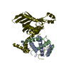
| ||||||||
|---|---|---|---|---|---|---|---|---|---|
| 1 | 
| ||||||||
| Unit cell |
| ||||||||
| Components on special symmetry positions |
|
-Components
| #1: Protein | Mass: 27294.443 Da / Num. of mol.: 1 Source method: isolated from a genetically manipulated source Source: (gene. exp.) Homo sapiens (human) / Gene: LDB1, CLIM2 / Production host:  Escherichia coli (E. coli) / References: UniProt: Q86U70 Escherichia coli (E. coli) / References: UniProt: Q86U70 | ||||
|---|---|---|---|---|---|
| #2: Protein | Mass: 10863.145 Da / Num. of mol.: 2 Source method: isolated from a genetically manipulated source Source: (gene. exp.) Homo sapiens (human) / Gene: SSBP2, SSDP2 / Production host: Escherichia coli (E. coli) / References: UniProt: P81877#3: Protein/peptide | | Mass: 2966.210 Da / Num. of mol.: 1 Source method: isolated from a genetically manipulated source Details: mutant 58Y was generated for monitoring UV280 to know where proteins are during purification Source: (gene. exp.) Homo sapiens (human) / Gene: PYGO2, PP7910 / Production host: Escherichia coli (E. coli) / References: UniProt: Q9BRQ0#4: Water | ChemComp-HOH / | Water Mass: 18.015 Da / Num. of mol.: 67 / Source method: isolated from a natural source / Formula: H2O Mass: 18.015 Da / Num. of mol.: 67 / Source method: isolated from a natural source / Formula: H2O |
-Experimental details
-Experiment
| Experiment | Method: X-RAY DIFFRACTION / Number of used crystals: 1 |
|---|
- Sample preparation
| Crystal | Density Matthews: 3.82 Å3/Da / Density % sol: 67.84 % |
|---|---|
| Crystal grow | Temperature: 298 K / Method: vapor diffusion, hanging drop Details: 100 mM Li2SO4, 100 mM sodium citrate tribasic dihydrate pH 5.6, 1% v/v PEG400, and 10 mM DTT |
-Data collection
| Diffraction | Mean temperature: 100 K / Serial crystal experiment: N |
|---|---|
| Diffraction source | Source: SYNCHROTRON / Site: NFPSS / Beamline: BL19U1 / Wavelength: 0.9792 Å |
| Detector | Type: DECTRIS PILATUS3 6M / Detector: PIXEL / Date: Jan 11, 2019 |
| Radiation | Protocol: SINGLE WAVELENGTH / Monochromatic (M) / Laue (L): M / Scattering type: x-ray |
| Radiation wavelength | Wavelength: 0.9792 Å / Relative weight: 1 |
| Reflection | Resolution: 2.45→20 Å / Num. obs: 30828 / % possible obs: 100 % / Redundancy: 12.7 % / CC1/2: 0.998 / Net I/σ(I): 44.86 |
| Reflection shell | Resolution: 2.45→2.52 Å / Num. unique obs: 2491 / CC1/2: 0.614 |
- Processing
| Software |
| |||||||||||||||||||||||||||||||||||||||||||||||||||||||||||||||||||||||||||||||||||||||||||||||||||||||||||||||||||||||||||||
|---|---|---|---|---|---|---|---|---|---|---|---|---|---|---|---|---|---|---|---|---|---|---|---|---|---|---|---|---|---|---|---|---|---|---|---|---|---|---|---|---|---|---|---|---|---|---|---|---|---|---|---|---|---|---|---|---|---|---|---|---|---|---|---|---|---|---|---|---|---|---|---|---|---|---|---|---|---|---|---|---|---|---|---|---|---|---|---|---|---|---|---|---|---|---|---|---|---|---|---|---|---|---|---|---|---|---|---|---|---|---|---|---|---|---|---|---|---|---|---|---|---|---|---|---|---|---|
| Refinement | Method to determine structure: MOLECULAR REPLACEMENT Starting model: 6TYD Resolution: 2.45→19.92 Å / SU ML: 0.36 / Cross valid method: NONE / σ(F): 1.35 / Phase error: 25.93 / Stereochemistry target values: ML
| |||||||||||||||||||||||||||||||||||||||||||||||||||||||||||||||||||||||||||||||||||||||||||||||||||||||||||||||||||||||||||||
| Solvent computation | Shrinkage radii: 0.9 Å / VDW probe radii: 1.11 Å / Solvent model: FLAT BULK SOLVENT MODEL | |||||||||||||||||||||||||||||||||||||||||||||||||||||||||||||||||||||||||||||||||||||||||||||||||||||||||||||||||||||||||||||
| Refinement step | Cycle: LAST / Resolution: 2.45→19.92 Å
| |||||||||||||||||||||||||||||||||||||||||||||||||||||||||||||||||||||||||||||||||||||||||||||||||||||||||||||||||||||||||||||
| Refine LS restraints |
| |||||||||||||||||||||||||||||||||||||||||||||||||||||||||||||||||||||||||||||||||||||||||||||||||||||||||||||||||||||||||||||
| LS refinement shell |
| |||||||||||||||||||||||||||||||||||||||||||||||||||||||||||||||||||||||||||||||||||||||||||||||||||||||||||||||||||||||||||||
| Refinement TLS params. | Method: refined / Refine-ID: X-RAY DIFFRACTION
| |||||||||||||||||||||||||||||||||||||||||||||||||||||||||||||||||||||||||||||||||||||||||||||||||||||||||||||||||||||||||||||
| Refinement TLS group |
|
