 Movie
Movie Controller


+ Open data

- Basic information
| Entry | Database: PDB / ID: 8hhh | ||||||
|---|---|---|---|---|---|---|---|
| Title | The bacterial divisome protein complex FtsB-FtsL-FtsQ | ||||||
 Components Components |
| ||||||
 Keywords Keywords |  MEMBRANE PROTEIN / Bacterial cell division / divisome / FtsB / FtsL / FtsQ / FtsBLQ / FtsQLB / membrane protein complex / heterotrimer MEMBRANE PROTEIN / Bacterial cell division / divisome / FtsB / FtsL / FtsQ / FtsBLQ / FtsQLB / membrane protein complex / heterotrimer | ||||||
| Function / homology |  Function and homology information Function and homology informationcell septum assembly / FtsZ-dependent cytokinesis / cell division site / plasma membraneSimilarity search - Function | ||||||
| Biological species |  Escherichia coli K-12 (bacteria) Escherichia coli K-12 (bacteria) | ||||||
| Method | X-RAY DIFFRACTION / SYNCHROTRON / MOLECULAR REPLACEMENT / Resolution: 3.3 Å | ||||||
 Authors Authors | Nguyen, V.H.T. / Chen, X. | ||||||
| Funding support |  Taiwan, 1items Taiwan, 1items
| ||||||
 Citation Citation | Journal: Nat Commun / Year: 2023 Title: Structure of the heterotrimeric membrane protein complex FtsB-FtsL-FtsQ of the bacterial divisome. Authors: Nguyen, H.T.V. / Chen, X. / Parada, C. / Luo, A.C. / Shih, O. / Jeng, U.S. / Huang, C.Y. / Shih, Y.L. / Ma, C. | ||||||
| History |
|
- Structure visualization
| Structure viewer | Molecule: MolmilJmol/JSmol |
|---|
- Downloads & links
-Download
| PDBx/mmCIF format | 8hhh.cif.gz | 184.2 KB | Display | PDBx/mmCIF format |
|---|---|---|---|---|
| PDB format | pdb8hhh.ent.gz | 146 KB | Display | PDB format |
| PDBx/mmJSON format | 8hhh.json.gz | Tree view | PDBx/mmJSON format | |
| Others |  Other downloads Other downloads |
-Validation report
| Arichive directory | https://data.pdbj.org/pub/pdb/validation_reports/hh/8hhhftp://data.pdbj.org/pub/pdb/validation_reports/hh/8hhh | HTTPS FTP |
|---|
-Related structure data
| Related structure data | 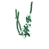 8hhfC  8hhgSC S: Starting model for refinement C: citing same article ( |
|---|---|
| Similar structure data |
-Links
 PDBj
PDBj- Assembly
| Deposited unit | 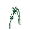
| ||||||||
|---|---|---|---|---|---|---|---|---|---|
| 1 |
| ||||||||
| Unit cell |
|
-Components
| #1: Protein | Mass: 31467.660 Da / Num. of mol.: 1 Source method: isolated from a genetically manipulated source Source: (gene. exp.) Escherichia coli K-12 (bacteria) / Strain: K-12Gene: ftsQ, A9X72_20810, ACU57_16060, BGM66_003029, BN17_45151, BO068_001919, BON68_06480, BON72_08970, BON73_04910, BON74_02170, BON75_16740, BON76_15060, BON77_21520, BON80_15910, BON89_27020, ...Gene: ftsQ, A9X72_20810, ACU57_16060, BGM66_003029, BN17_45151, BO068_001919, BON68_06480, BON72_08970, BON73_04910, BON74_02170, BON75_16740, BON76_15060, BON77_21520, BON80_15910, BON89_27020, BON93_20085, BON94_26730, BvCmsHHP019_03716, C5Y87_12070, CCS08_22990, CR538_21045, CTR35_001627, DAH19_13315, DAH50_07465, DS732_05370, DTL43_06330, DXT70_08670, E2119_07785, E2134_11600, E4K51_06165, E5P23_04335, E5P31_03955, E5P32_08015, E5P35_00730, E5P36_04260, E5P39_13665, E5P40_07375, E5P41_14185, E5P42_01010, E5P43_09200, E5P44_09730, E5P45_08355, E5P46_13085, E5P47_04370, E5P48_12150, E5P49_11070, E5P50_01390, E5S36_10750, E5S51_07905, EI021_04715, EIZ93_07570, EL79_3779, EL80_3726, ELT20_04095, ELT41_01600, ELX85_12445, F2N20_00540, F2N31_00540, F9V24_01135, FJQ40_06395, FOI11_013215, FOI11_22465, FV293_03270, FWK02_17275, GJO56_09885, GKF89_02115, GNW61_10395, GP944_03500, GP965_07730, GP979_06755, GRW05_06695, HMV95_04965, HX136_20990, IH768_05560, J0541_002403, JNP96_04660, NCTC11126_03516, NCTC13216_02643, NCTC8008_03748, NCTC8500_04640, NCTC9037_04264, NCTC9045_04807, NCTC9117_05152, NCTC9706_01498, SAMEA3751407_02594, SAMEA3752557_00798 Production host: Escherichia coli (E. coli) / References: UniProt: J7Q602 |
|---|---|
| #2: Protein | Mass: 13397.883 Da / Num. of mol.: 1 / Mutation: E56A Source method: isolated from a genetically manipulated source Source: (gene. exp.) Escherichia coli K-12 (bacteria) / Strain: K-12 / Gene: ygbQ, ftsB / Production host: Escherichia coli (E. coli) / References: UniProt: Q1JQN6 |
| #3: Protein | Mass: 13646.827 Da / Num. of mol.: 1 Source method: isolated from a genetically manipulated source Source: (gene. exp.) Escherichia coli K-12 (bacteria) / Strain: K-12 / Gene: ftsL / Production host: Escherichia coli (E. coli) / References: UniProt: D6II44 |
-Experimental details
-Experiment
| Experiment | Method: X-RAY DIFFRACTION / Number of used crystals: 1 |
|---|
- Sample preparation
| Crystal | Density Matthews: 4.31 Å3/Da / Density % sol: 71.49 % |
|---|---|
| Crystal grow | Temperature: 293 K / Method: vapor diffusion, sitting drop / pH: 8 / Details: 0.1 M Tris pH 8.0, 20-35% PEG400 |
-Data collection
| Diffraction | Mean temperature: 100 K / Serial crystal experiment: N |
|---|---|
| Diffraction source | Source: SYNCHROTRON / Site: NSRRC / Beamline: TPS 05A / Wavelength: 1 Å |
| Detector | Type: RAYONIX MX300-HS / Detector: CCD / Date: Dec 22, 2020 |
| Radiation | Monochromator: liquid-nitrogen-cooling Si double-crystal / Protocol: SINGLE WAVELENGTH / Monochromatic (M) / Laue (L): M / Scattering type: x-ray |
| Radiation wavelength | Wavelength: 1 Å / Relative weight: 1 |
| Reflection | Resolution: 3.3→90.33 Å / Num. obs: 14819 / % possible obs: 99.83 % / Redundancy: 3.7 % / Rmerge(I) obs: 0.07429 / Net I/σ(I): 9.25 |
| Reflection shell | Resolution: 3.3→3.56 Å / Rmerge(I) obs: 0.6163 / Num. unique obs: 1476 |
- Processing
| Software |
| ||||||||||||||||||||||||||||||||||||||||
|---|---|---|---|---|---|---|---|---|---|---|---|---|---|---|---|---|---|---|---|---|---|---|---|---|---|---|---|---|---|---|---|---|---|---|---|---|---|---|---|---|---|
| Refinement | Method to determine structure: MOLECULAR REPLACEMENT Starting model: 8HHG Resolution: 3.3→34.69 Å / SU ML: 0.48 / Cross valid method: THROUGHOUT / σ(F): 1.36 / Phase error: 39 / Stereochemistry target values: ML
| ||||||||||||||||||||||||||||||||||||||||
| Solvent computation | Shrinkage radii: 0.9 Å / VDW probe radii: 1.11 Å / Solvent model: FLAT BULK SOLVENT MODEL | ||||||||||||||||||||||||||||||||||||||||
| Displacement parameters | Biso max: 279.05 Å2 / Biso mean: 127.6639 Å2 / Biso min: 30 Å2 | ||||||||||||||||||||||||||||||||||||||||
| Refinement step | Cycle: final / Resolution: 3.3→34.69 Å
| ||||||||||||||||||||||||||||||||||||||||
| LS refinement shell | Refine-ID: X-RAY DIFFRACTION / Rfactor Rfree error: 0 / % reflection obs: 100 %
| ||||||||||||||||||||||||||||||||||||||||
| Refinement TLS params. | Method: refined / Origin x: -17.5432 Å / Origin y: -4.0766 Å / Origin z: -16.0345 Å
| ||||||||||||||||||||||||||||||||||||||||
| Refinement TLS group |
|
