 Movie
Movie Controller


+ Open data

- Basic information
| Entry | Database: PDB / ID: 8h5z | ||||||
|---|---|---|---|---|---|---|---|
| Title | Crystal structure of RadD/ATP analogue complex | ||||||
 Components Components | Putative DNA repair helicase RadD | ||||||
 Keywords Keywords |  DNA BINDING PROTEIN / helicase superfamily 2 DNA repair ATPase DNA BINDING PROTEIN / helicase superfamily 2 DNA repair ATPase | ||||||
| Function / homology |  Function and homology informationpostreplication repair / response to ionizing radiation / helicase activity / double-strand break repair / single-stranded DNA binding / DNA helicase / response to xenobiotic stimulus / translation / cell division / ATP hydrolysis activity ...postreplication repair / response to ionizing radiation / helicase activity / double-strand break repair / single-stranded DNA binding / DNA helicase / response to xenobiotic stimulus / translation / cell division / ATP hydrolysis activity / DNA binding / zinc ion binding / ATP binding Function and homology informationpostreplication repair / response to ionizing radiation / helicase activity / double-strand break repair / single-stranded DNA binding / DNA helicase / response to xenobiotic stimulus / translation / cell division / ATP hydrolysis activity ...postreplication repair / response to ionizing radiation / helicase activity / double-strand break repair / single-stranded DNA binding / DNA helicase / response to xenobiotic stimulus / translation / cell division / ATP hydrolysis activity / DNA binding / zinc ion binding / ATP bindingSimilarity search - Function | ||||||
| Biological species |  Escherichia coli K-12 (bacteria) Escherichia coli K-12 (bacteria) | ||||||
| Method | X-RAY DIFFRACTION / SYNCHROTRON / MOLECULAR REPLACEMENT / Resolution: 3.00002725136 Å | ||||||
 Authors Authors | Yan, X.X. / Tian, L.F. | ||||||
| Funding support | 1items
| ||||||
 Citation Citation | Journal: Int J Mol Sci / Year: 2023 Title: Biochemical and Structural Analyses Shed Light on the Mechanisms of RadD DNA Binding and Its ATPase from Escherichia coli. Authors: Tian, L.F. / Kuang, X. / Ding, K. / Gao, H. / Tang, Q. / Yan, X.X. / Xu, W. | ||||||
| History |
|
- Structure visualization
| Structure viewer | Molecule: MolmilJmol/JSmol |
|---|
- Downloads & links
-Download
| PDBx/mmCIF format | 8h5z.cif.gz | 232.3 KB | Display | PDBx/mmCIF format |
|---|---|---|---|---|
| PDB format | pdb8h5z.ent.gz | 181.7 KB | Display | PDB format |
| PDBx/mmJSON format | 8h5z.json.gz | Tree view | PDBx/mmJSON format | |
| Others |  Other downloads Other downloads |
-Validation report
| Arichive directory | https://data.pdbj.org/pub/pdb/validation_reports/h5/8h5zftp://data.pdbj.org/pub/pdb/validation_reports/h5/8h5z | HTTPS FTP |
|---|
-Related structure data
| Related structure data |  8h5yC 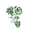 6jdeS S: Starting model for refinement C: citing same article ( |
|---|---|
| Similar structure data |
-Links
 PDBj
PDBj
- Assembly
| Deposited unit | 
| ||||||||||||
|---|---|---|---|---|---|---|---|---|---|---|---|---|---|
| 1 |
| ||||||||||||
| 2 |
| ||||||||||||
| Unit cell |
|
-Components
| #1: Protein | Mass: 66509.039 Da / Num. of mol.: 2 Source method: isolated from a genetically manipulated source Source: (gene. exp.) Escherichia coli K-12 (bacteria) / Gene: radD, yejH, yejI, yejJ, b2184, JW2172 / Production host: Escherichia coli BL21 (bacteria) / References: UniProt: P33919, DNA helicase#2: Chemical |   Mass: 523.247 Da / Num. of mol.: 2 / Source method: obtained synthetically / Formula: C10H16N5O12P3S / Feature type: SUBJECT OF INVESTIGATION / Comment: ATP-gamma-S, energy-carrying molecule analogue*YM Mass: 523.247 Da / Num. of mol.: 2 / Source method: obtained synthetically / Formula: C10H16N5O12P3S / Feature type: SUBJECT OF INVESTIGATION / Comment: ATP-gamma-S, energy-carrying molecule analogue*YM#3: Chemical |   Mass: 65.409 Da / Num. of mol.: 2 / Source method: obtained synthetically / Formula: Zn / Feature type: SUBJECT OF INVESTIGATION Mass: 65.409 Da / Num. of mol.: 2 / Source method: obtained synthetically / Formula: Zn / Feature type: SUBJECT OF INVESTIGATIONHas ligand of interest | Y | |
|---|
-Experimental details
-Experiment
| Experiment | Method: X-RAY DIFFRACTION / Number of used crystals: 1 |
|---|
- Sample preparation
| Crystal | Density Matthews: 2.66 Å3/Da / Density % sol: 53.75 % |
|---|---|
| Crystal grow | Temperature: 286 K / Method: vapor diffusion, hanging drop / Details: 500 mM NaCl, 5 mM MgCl2 and 18% [w/v] PEG3350 |
-Data collection
| Diffraction | Mean temperature: 100 K / Serial crystal experiment: N |
|---|---|
| Diffraction source | Source: SYNCHROTRON / Site: SSRF  / Beamline: BL19U1 / Wavelength: 0.97929 Å / Beamline: BL19U1 / Wavelength: 0.97929 Å |
| Detector | Type: DECTRIS PILATUS3 6M / Detector: PIXEL / Date: Jul 12, 2018 |
| Radiation | Protocol: SINGLE WAVELENGTH / Monochromatic (M) / Laue (L): M / Scattering type: x-ray |
| Radiation wavelength | Wavelength: 0.97929 Å / Relative weight: 1 |
| Reflection | Resolution: 3→20 Å / Num. obs: 38570 / % possible obs: 99.8 % / Redundancy: 6.5 % / Biso Wilson estimate: 68.6097829319 Å2 / CC1/2: 0.795 / Net I/σ(I): 14.3 |
| Reflection shell | Resolution: 3→3.12 Å / Num. unique obs: 1910 / CC1/2: 0.795 |
- Processing
| Software |
| |||||||||||||||||||||||||||||||||||||||||||||||||||||||||||||||||||||||||||||
|---|---|---|---|---|---|---|---|---|---|---|---|---|---|---|---|---|---|---|---|---|---|---|---|---|---|---|---|---|---|---|---|---|---|---|---|---|---|---|---|---|---|---|---|---|---|---|---|---|---|---|---|---|---|---|---|---|---|---|---|---|---|---|---|---|---|---|---|---|---|---|---|---|---|---|---|---|---|---|
| Refinement | Method to determine structure: MOLECULAR REPLACEMENT Starting model: 6JDE Resolution: 3.00002725136→19.9761477285 Å / SU ML: 0.417256574005 / Cross valid method: FREE R-VALUE / σ(F): 1.35546330902 / Phase error: 33.7639508679 Stereochemistry target values: GeoStd + Monomer Library + CDL v1.2
| |||||||||||||||||||||||||||||||||||||||||||||||||||||||||||||||||||||||||||||
| Solvent computation | Shrinkage radii: 0.9 Å / VDW probe radii: 1.11 Å / Solvent model: FLAT BULK SOLVENT MODEL | |||||||||||||||||||||||||||||||||||||||||||||||||||||||||||||||||||||||||||||
| Displacement parameters | Biso mean: 65.9507554787 Å2 | |||||||||||||||||||||||||||||||||||||||||||||||||||||||||||||||||||||||||||||
| Refinement step | Cycle: LAST / Resolution: 3.00002725136→19.9761477285 Å
| |||||||||||||||||||||||||||||||||||||||||||||||||||||||||||||||||||||||||||||
| Refine LS restraints |
| |||||||||||||||||||||||||||||||||||||||||||||||||||||||||||||||||||||||||||||
| LS refinement shell |
|
