 Movie
Movie Controller

[English] 日本語
 Yorodumi
- PDB-8gyr: Crystal structure of a variable region segment of Leptospira host... -
+ Open data

- Basic information
| Entry | Database: PDB / ID: 8gyr | ||||||
|---|---|---|---|---|---|---|---|
| Title | Crystal structure of a variable region segment of Leptospira host-interacting outer surface protein, LigA | ||||||
 Components Components |
| ||||||
 Keywords Keywords |  CELL ADHESION / Leptospira / Leptospira immunoglobulin-like protein (Lig) / microbial surface components recognizing adhesive matrix molecule (MSCRAMM) CELL ADHESION / Leptospira / Leptospira immunoglobulin-like protein (Lig) / microbial surface components recognizing adhesive matrix molecule (MSCRAMM) | ||||||
| Function / homology | Invasin/intimin cell-adhesion fragments / Bacterial Ig-like domain (group 2) / Bacterial Ig-like domain 2 / Bacterial Ig-like domain, group 2 / Prokaryotic membrane lipoprotein lipid attachment site profile. / IODIDE ION / : / PHOSPHATE ION / LigA Function and homology information Function and homology information | ||||||
| Biological species |  Leptospira interrogans (bacteria) Leptospira interrogans (bacteria) | ||||||
| Method | X-RAY DIFFRACTION / MOLECULAR REPLACEMENT / Resolution: 1.87 Å | ||||||
 Authors Authors | Kumar, P. / Akif, M. | ||||||
| Funding support |  India, 1items India, 1items
| ||||||
 Citation Citation | Journal: Int.J.Biol.Macromol. / Year: 2023 Title: Crystal structure of a variable region segment of Leptospira host-interacting outer surface protein, LigA, reveals the orientation of Ig-like domains. Authors: Kumar, P. / Vyas, P. / Faisal, S.M. / Chang, Y.F. / Akif, M. | ||||||
| History |
|
- Structure visualization
| Structure viewer | Molecule: MolmilJmol/JSmol |
|---|
- Downloads & links
-Download
| PDBx/mmCIF format | 8gyr.cif.gz | 155.9 KB | Display | PDBx/mmCIF format |
|---|---|---|---|---|
| PDB format | pdb8gyr.ent.gz | 120 KB | Display | PDB format |
| PDBx/mmJSON format | 8gyr.json.gz | Tree view | PDBx/mmJSON format | |
| Others |  Other downloads Other downloads |
-Validation report
| Arichive directory | https://data.pdbj.org/pub/pdb/validation_reports/gy/8gyrftp://data.pdbj.org/pub/pdb/validation_reports/gy/8gyr | HTTPS FTP |
|---|
-Related structure data
| Related structure data | 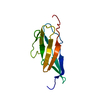 2mogS S: Starting model for refinement |
|---|---|
| Similar structure data |
-Links
 PDBj
PDBj


- Assembly
| Deposited unit | 
| ||||||||
|---|---|---|---|---|---|---|---|---|---|
| 1 | 
| ||||||||
| 2 | 
| ||||||||
| Unit cell |
|
-Components
-Protein , 2 types, 2 molecules AB
| #1: Protein | Mass: 18811.148 Da / Num. of mol.: 1 Source method: isolated from a genetically manipulated source Source: (gene. exp.) Leptospira interrogans (bacteria) / Production host: Escherichia coli (E. coli) / References: UniProt: C0J1Q2 |
|---|---|
| #2: Protein | Mass: 18967.289 Da / Num. of mol.: 1 Source method: isolated from a genetically manipulated source Source: (gene. exp.) Leptospira interrogans (bacteria) / Gene: ligA / Production host: Escherichia coli (E. coli) / References: UniProt: C0J1Q2 |
-Non-polymers , 8 types, 336 molecules 


| #3: Chemical | ChemComp-K /  Mass: 39.098 Da / Num. of mol.: 5 / Source method: obtained synthetically / Formula: K Mass: 39.098 Da / Num. of mol.: 5 / Source method: obtained synthetically / Formula: K#4: Chemical | ChemComp-CL / | Chloride Mass: 35.453 Da / Num. of mol.: 1 / Source method: obtained synthetically / Formula: Cl Mass: 35.453 Da / Num. of mol.: 1 / Source method: obtained synthetically / Formula: Cl#5: Chemical | ChemComp-EDO / Ethylene glycol Mass: 62.068 Da / Num. of mol.: 4 / Source method: obtained synthetically / Formula: C2H6O2 Mass: 62.068 Da / Num. of mol.: 4 / Source method: obtained synthetically / Formula: C2H6O2#6: Chemical | Phosphate Mass: 94.971 Da / Num. of mol.: 2 / Source method: obtained synthetically / Formula: PO4 Mass: 94.971 Da / Num. of mol.: 2 / Source method: obtained synthetically / Formula: PO4#7: Chemical | ChemComp-NA /  Mass: 22.990 Da / Num. of mol.: 5 / Source method: obtained synthetically / Formula: Na Mass: 22.990 Da / Num. of mol.: 5 / Source method: obtained synthetically / Formula: Na#8: Chemical |  Mass: 40.078 Da / Num. of mol.: 2 / Source method: obtained synthetically / Formula: Ca Mass: 40.078 Da / Num. of mol.: 2 / Source method: obtained synthetically / Formula: Ca#9: Chemical | Iodide Mass: 126.904 Da / Num. of mol.: 2 / Source method: obtained synthetically / Formula: I Mass: 126.904 Da / Num. of mol.: 2 / Source method: obtained synthetically / Formula: I#10: Water | ChemComp-HOH / | WaterMass: 18.015 Da / Num. of mol.: 315 / Source method: isolated from a natural source / Formula: H2O |
|---|
-Details
| Has ligand of interest | N |
|---|
-Experimental details
-Experiment
| Experiment | Method: X-RAY DIFFRACTION / Number of used crystals: 1 |
|---|
- Sample preparation
| Crystal | Density Matthews: 2.45 Å3/Da / Density % sol: 49.86 % |
|---|---|
| Crystal grow | Temperature: 295 K / Method: vapor diffusion, hanging drop / pH: 7.5 Details: 23% PEG 3350, 260mM Potassium Iodide and 100mM Bis-Tris Propane |
-Data collection
| Diffraction | Mean temperature: 100 K / Serial crystal experiment: N |
|---|---|
| Diffraction source | Source: ROTATING ANODE / Type: RIGAKU MICROMAX-007 HF / Wavelength: 1.5418 Å |
| Detector | Type: MAR scanner 345 mm plate / Detector: IMAGE PLATE / Date: Mar 21, 2019 |
| Radiation | Protocol: SINGLE WAVELENGTH / Monochromatic (M) / Laue (L): M / Scattering type: x-ray |
| Radiation wavelength | Wavelength: 1.5418 Å / Relative weight: 1 |
| Reflection | Resolution: 1.87→85.92 Å / Num. obs: 31943 / % possible obs: 98.8 % / Redundancy: 6.7 % / CC1/2: 0.994 / Rmerge(I) obs: 0.113 / Net I/σ(I): 9.1 |
| Reflection shell | Resolution: 1.87→1.91 Å / Num. unique obs: 1705 / CC1/2: 0.759 |
- Processing
| Software |
| |||||||||||||||||||||||||||||||||||||||||||||||||||||||||||||||||||||||||||||||||||||||||||||||||||||||||
|---|---|---|---|---|---|---|---|---|---|---|---|---|---|---|---|---|---|---|---|---|---|---|---|---|---|---|---|---|---|---|---|---|---|---|---|---|---|---|---|---|---|---|---|---|---|---|---|---|---|---|---|---|---|---|---|---|---|---|---|---|---|---|---|---|---|---|---|---|---|---|---|---|---|---|---|---|---|---|---|---|---|---|---|---|---|---|---|---|---|---|---|---|---|---|---|---|---|---|---|---|---|---|---|---|---|---|
| Refinement | Method to determine structure: MOLECULAR REPLACEMENT Starting model: 2MOG Resolution: 1.87→85.92 Å / Cor.coef. Fo:Fc: 0.93 / Cor.coef. Fo:Fc free: 0.906 / SU B: 7.551 / SU ML: 0.118 / Cross valid method: THROUGHOUT / ESU R: 0.161 / ESU R Free: 0.141 / Stereochemistry target values: MAXIMUM LIKELIHOOD Details: U VALUES : WITH TLS ADDED HYDROGENS HAVE BEEN ADDED IN THE RIDING POSITIONS U VALUES : RESIDUAL ONLY
| |||||||||||||||||||||||||||||||||||||||||||||||||||||||||||||||||||||||||||||||||||||||||||||||||||||||||
| Solvent computation | Ion probe radii: 1 Å / Shrinkage radii: 1 Å / VDW probe radii: 1.3 Å / Solvent model: MASK | |||||||||||||||||||||||||||||||||||||||||||||||||||||||||||||||||||||||||||||||||||||||||||||||||||||||||
| Displacement parameters | Biso mean: 21.308 Å2
| |||||||||||||||||||||||||||||||||||||||||||||||||||||||||||||||||||||||||||||||||||||||||||||||||||||||||
| Refinement step | Cycle: LAST / Resolution: 1.87→85.92 Å
| |||||||||||||||||||||||||||||||||||||||||||||||||||||||||||||||||||||||||||||||||||||||||||||||||||||||||
| Refine LS restraints |
| |||||||||||||||||||||||||||||||||||||||||||||||||||||||||||||||||||||||||||||||||||||||||||||||||||||||||
| LS refinement shell | Resolution: 1.87→1.917 Å
| |||||||||||||||||||||||||||||||||||||||||||||||||||||||||||||||||||||||||||||||||||||||||||||||||||||||||
| Refinement TLS params. | Method: refined / Refine-ID: X-RAY DIFFRACTION
| |||||||||||||||||||||||||||||||||||||||||||||||||||||||||||||||||||||||||||||||||||||||||||||||||||||||||
| Refinement TLS group |
|
