 Movie
Movie Controller


+ Open data

- Basic information
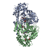
| Entry | Database: PDB / ID: 8gyh | |||||||||||||||
|---|---|---|---|---|---|---|---|---|---|---|---|---|---|---|---|---|
| Title | Crystal structure of Fic25 (apo form) from Streptomyces ficellus | |||||||||||||||
 Components Components | DegT/DnrJ/EryC1/StrS family aminotransferase | |||||||||||||||
 Keywords Keywords |  BIOSYNTHETIC PROTEIN / Sugar aminotransferase / Biosynthetic enzyme / DADH BIOSYNTHETIC PROTEIN / Sugar aminotransferase / Biosynthetic enzyme / DADH | |||||||||||||||
| Function / homology |  Function and homology informationdTDP-4-amino-4,6-dideoxygalactose transaminase activity / polysaccharide biosynthetic process / antibiotic biosynthetic process / pyridoxal phosphate binding Function and homology informationdTDP-4-amino-4,6-dideoxygalactose transaminase activity / polysaccharide biosynthetic process / antibiotic biosynthetic process / pyridoxal phosphate bindingSimilarity search - Function | |||||||||||||||
| Biological species |  Streptomyces ficellus (bacteria) Streptomyces ficellus (bacteria) | |||||||||||||||
| Method | X-RAY DIFFRACTION / SYNCHROTRON / MOLECULAR REPLACEMENT / Resolution: 1.8 Å | |||||||||||||||
 Authors Authors | Kurosawa, S. / Yoshida, A. / Tomita, T. / Nishiyama, M. | |||||||||||||||
| Funding support |  Japan, 4items Japan, 4items
| |||||||||||||||
 Citation Citation | Journal: Acs Chem.Biol. / Year: 2023 Title: Mechanisms of Sugar Aminotransferase-like Enzymes to Synthesize Stereoisomers of Non-proteinogenic Amino Acids in Natural Product Biosynthesis. Authors: Kurosawa, S. / Okamura, H. / Yoshida, A. / Tomita, T. / Sone, Y. / Hasebe, F. / Shinada, T. / Takikawa, H. / Kosono, S. / Nishiyama, M. | |||||||||||||||
| History |
|
- Structure visualization
| Structure viewer | Molecule: MolmilJmol/JSmol |
|---|
- Downloads & links
-Download
| PDBx/mmCIF format | 8gyh.cif.gz | 188.1 KB | Display | PDBx/mmCIF format |
|---|---|---|---|---|
| PDB format | pdb8gyh.ent.gz | 147.4 KB | Display | PDB format |
| PDBx/mmJSON format | 8gyh.json.gz | Tree view | PDBx/mmJSON format | |
| Others |  Other downloads Other downloads |
-Validation report
| Arichive directory | https://data.pdbj.org/pub/pdb/validation_reports/gy/8gyhftp://data.pdbj.org/pub/pdb/validation_reports/gy/8gyh | HTTPS FTP |
|---|
-Related structure data

-Links
 PDBj
PDBj


- Assembly
| Deposited unit | 
| ||||||||
|---|---|---|---|---|---|---|---|---|---|
| 1 |
| ||||||||
| Unit cell |
|
-Components
| #1: Protein | Mass: 46911.789 Da / Num. of mol.: 2 Source method: isolated from a genetically manipulated source Source: (gene. exp.) Streptomyces ficellus (bacteria) / Gene: EIZ62_06105 / Production host: Escherichia coli BL21(DE3) (bacteria) / References: UniProt: A0A1W5T2G9#2: Chemical | ChemComp-GOL / | Glycerol  Mass: 92.094 Da / Num. of mol.: 1 / Source method: obtained synthetically / Formula: C3H8O3 Mass: 92.094 Da / Num. of mol.: 1 / Source method: obtained synthetically / Formula: C3H8O3#3: Chemical | ChemComp-IMD / Imidazole  Mass: 69.085 Da / Num. of mol.: 8 / Source method: obtained synthetically / Formula: C3H5N2 Mass: 69.085 Da / Num. of mol.: 8 / Source method: obtained synthetically / Formula: C3H5N2#4: Water | ChemComp-HOH / | Water Mass: 18.015 Da / Num. of mol.: 745 / Source method: isolated from a natural source / Formula: H2O Mass: 18.015 Da / Num. of mol.: 745 / Source method: isolated from a natural source / Formula: H2OHas ligand of interest | N | |
|---|
-Experimental details
-Experiment
| Experiment | Method: X-RAY DIFFRACTION / Number of used crystals: 1 |
|---|
- Sample preparation
| Crystal | Density Matthews: 3.57 Å3/Da / Density % sol: 65.5 % |
|---|---|
| Crystal grow | Temperature: 293.15 K / Method: vapor diffusion, hanging drop / Details: 1.0 M Sodium citrate, 0.1 M Imidazole, pH8.0 |
-Data collection
| Diffraction | Mean temperature: 95 K / Serial crystal experiment: N |
|---|---|
| Diffraction source | Source: SYNCHROTRON / Site: Photon Factory / Beamline: BL-5A / Wavelength: 1 Å |
| Detector | Type: DECTRIS PILATUS3 6M / Detector: PIXEL / Date: Nov 27, 2021 |
| Radiation | Protocol: SINGLE WAVELENGTH / Monochromatic (M) / Laue (L): M / Scattering type: x-ray |
| Radiation wavelength | Wavelength: 1 Å / Relative weight: 1 |
| Reflection | Resolution: 1.8→48.484 Å / Num. obs: 122992 / % possible obs: 100 % / Redundancy: 10.2 % / CC1/2: 0.999 / Rmerge(I) obs: 0.112 / Net I/σ(I): 18.9 |
| Reflection shell | Resolution: 1.8→1.83 Å / Rmerge(I) obs: 0.94 / Num. unique obs: 6059 / CC1/2: 0.835 |
- Processing
| Software |
| |||||||||||||||||||||||||||||||||||||||||||||||||||||||
|---|---|---|---|---|---|---|---|---|---|---|---|---|---|---|---|---|---|---|---|---|---|---|---|---|---|---|---|---|---|---|---|---|---|---|---|---|---|---|---|---|---|---|---|---|---|---|---|---|---|---|---|---|---|---|---|---|
| Refinement | Method to determine structure: MOLECULAR REPLACEMENT Starting model: Model constructed by AlphaFold2 from the primary amino acid sequence of Fic25 Resolution: 1.8→48.48 Å / Cor.coef. Fo:Fc: 0.962 / Cor.coef. Fo:Fc free: 0.953 / SU B: 2.223 / SU ML: 0.066 / Cross valid method: THROUGHOUT / σ(F): 0 / ESU R: 0.09 / ESU R Free: 0.087 / Stereochemistry target values: MAXIMUM LIKELIHOOD Details: HYDROGENS HAVE BEEN ADDED IN THE RIDING POSITIONS U VALUES : REFINED INDIVIDUALLY
| |||||||||||||||||||||||||||||||||||||||||||||||||||||||
| Solvent computation | Ion probe radii: 0.8 Å / Shrinkage radii: 0.8 Å / VDW probe radii: 1.2 Å / Solvent model: MASK | |||||||||||||||||||||||||||||||||||||||||||||||||||||||
| Displacement parameters | Biso max: 83.56 Å2 / Biso mean: 21.536 Å2 / Biso min: 11.7 Å2
| |||||||||||||||||||||||||||||||||||||||||||||||||||||||
| Refinement step | Cycle: final / Resolution: 1.8→48.48 Å
| |||||||||||||||||||||||||||||||||||||||||||||||||||||||
| Refine LS restraints |
| |||||||||||||||||||||||||||||||||||||||||||||||||||||||
| LS refinement shell | Resolution: 1.8→1.847 Å / Rfactor Rfree error: 0 / Total num. of bins used: 20
|
