 Movie
Movie Controller

[English] 日本語
 Yorodumi
- PDB-8grj: Crystal structure of gamma-alpha subunit complex from Burkholderi... -
+ Open data

- Basic information
| Entry | Database: PDB / ID: 8grj | ||||||
|---|---|---|---|---|---|---|---|
| Title | Crystal structure of gamma-alpha subunit complex from Burkholderia cepacia FAD glucose dehydrogenase in complex with gluconolactone | ||||||
 Components Components |
| ||||||
 Keywords Keywords |  OXIDOREDUCTASE / Glucose dehydrogenase / FAD / Burkholderia cepacia / SIGNALING PROTEIN-OXIDOREDUCTASE complex OXIDOREDUCTASE / Glucose dehydrogenase / FAD / Burkholderia cepacia / SIGNALING PROTEIN-OXIDOREDUCTASE complex | ||||||
| Function / homology |  Function and homology information Function and homology informationoxidoreductase activity, acting on CH-OH group of donors / 3 iron, 4 sulfur cluster binding / flavin adenine dinucleotide binding / metal ion bindingSimilarity search - Function | ||||||
| Biological species |  Burkholderia cepacia (bacteria) Burkholderia cepacia (bacteria) | ||||||
| Method | X-RAY DIFFRACTION / SYNCHROTRON / MOLECULAR REPLACEMENT / Resolution: 2.95 Å | ||||||
 Authors Authors | Yoshida, H. / Kojima, K. / Tsugawa, W. / Okuda-Shimazaki, J. / Kerrigan, J.A. / Sode, K. | ||||||
| Funding support |  Japan, 1items Japan, 1items
| ||||||
 Citation Citation | Journal: To Be Published Title: Crystal structure of gamma-alpha subunit complex from Burkholderia cepacia FAD glucose dehydrogenase in complex with gluconolactone Authors: Yoshida, H. / Kojima, K. / Tsugawa, W. / Okuda-Shimazaki, J. / Kerrigan, J.A. / Sode, K. #1: Journal: Acta Crystallogr D Struct Biol / Year: 2019Title: X-ray structure of the direct electron transfer-type FAD glucose dehydrogenase catalytic subunit complexed with a hitchhiker protein. Authors: Yoshida, H. / Kojima, K. / Shiota, M. / Yoshimatsu, K. / Yamazaki, T. / Ferri, S. / Tsugawa, W. / Kamitori, S. / Sode, K. | ||||||
| History |
|
- Structure visualization
| Structure viewer | Molecule: MolmilJmol/JSmol |
|---|
- Downloads & links
-Download
| PDBx/mmCIF format | 8grj.cif.gz | 267.8 KB | Display | PDBx/mmCIF format |
|---|---|---|---|---|
| PDB format | pdb8grj.ent.gz | 214 KB | Display | PDB format |
| PDBx/mmJSON format | 8grj.json.gz | Tree view | PDBx/mmJSON format | |
| Others |  Other downloads Other downloads |
-Validation report
| Arichive directory | https://data.pdbj.org/pub/pdb/validation_reports/gr/8grjftp://data.pdbj.org/pub/pdb/validation_reports/gr/8grj | HTTPS FTP |
|---|
-Related structure data
| Related structure data | 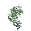 6a2uS S: Starting model for refinement |
|---|---|
| Similar structure data |
-Links
 PDBj
PDBj


- Assembly
| Deposited unit | 
| ||||||||
|---|---|---|---|---|---|---|---|---|---|
| 1 |
| ||||||||
| 2 |
| ||||||||
| Unit cell |
|
-Components
-Protein , 2 types, 4 molecules ACBD
| #1: Protein | Mass: 17980.414 Da / Num. of mol.: 2 Source method: isolated from a genetically manipulated source Details: GB:CAZ78686.1 / Source: (gene. exp.) Burkholderia cepacia (bacteria) / Strain: ATCC 17616 / 249 / Gene: BMULJ_04411 / Plasmid: pTrc99A / Production host: Escherichia coli BL21(DE3) (bacteria) / Strain (production host): BL21(DE3) / References: UniProt: A0A0H3KLY3#2: Protein | Mass: 60735.762 Da / Num. of mol.: 2 Source method: isolated from a genetically manipulated source Details: GB:AAN39686.1 / Source: (gene. exp.) Burkholderia cepacia (bacteria) / Gene: gdhAlpha / Plasmid: pTrc99A / Production host: Escherichia coli BL21(DE3) (bacteria) / Strain (production host): BL21(DE3) / References: UniProt: Q8GQE7 |
|---|
-Sugars , 1 types, 2 molecules 
| #5: Sugar | Glucono delta-lactone Type: D-saccharide / Mass: 178.140 Da / Num. of mol.: 2 / Source method: obtained synthetically / Formula: C6H10O6 / Feature type: SUBJECT OF INVESTIGATION Type: D-saccharide / Mass: 178.140 Da / Num. of mol.: 2 / Source method: obtained synthetically / Formula: C6H10O6 / Feature type: SUBJECT OF INVESTIGATION |
|---|
-Non-polymers , 3 types, 153 molecules 


| #3: Chemical | Flavin adenine dinucleotide Mass: 785.550 Da / Num. of mol.: 2 / Source method: obtained synthetically / Formula: C27H33N9O15P2 / Comment: FAD*YM Mass: 785.550 Da / Num. of mol.: 2 / Source method: obtained synthetically / Formula: C27H33N9O15P2 / Comment: FAD*YM#4: Chemical | Iron–sulfur cluster Mass: 295.795 Da / Num. of mol.: 2 / Source method: obtained synthetically / Formula: Fe3S4 Mass: 295.795 Da / Num. of mol.: 2 / Source method: obtained synthetically / Formula: Fe3S4#6: Water | ChemComp-HOH / | WaterMass: 18.015 Da / Num. of mol.: 149 / Source method: isolated from a natural source / Formula: H2O |
|---|
-Details
| Has ligand of interest | Y |
|---|
-Experimental details
-Experiment
| Experiment | Method: X-RAY DIFFRACTION / Number of used crystals: 1 |
|---|
- Sample preparation
| Crystal | Density Matthews: 2.99 Å3/Da / Density % sol: 58.86 % |
|---|---|
| Crystal grow | Temperature: 293 K / Method: vapor diffusion, sitting drop / pH: 7 / Details: Tacsimate |
-Data collection
| Diffraction | Mean temperature: 100 K / Serial crystal experiment: N | |||||||||||||||||||||||||||||||||||||||||||||||||||||||||||||||||||||||||||||||||||||||||||||||||||||||||||||||||||||||||||||||||||||||||||||||||||
|---|---|---|---|---|---|---|---|---|---|---|---|---|---|---|---|---|---|---|---|---|---|---|---|---|---|---|---|---|---|---|---|---|---|---|---|---|---|---|---|---|---|---|---|---|---|---|---|---|---|---|---|---|---|---|---|---|---|---|---|---|---|---|---|---|---|---|---|---|---|---|---|---|---|---|---|---|---|---|---|---|---|---|---|---|---|---|---|---|---|---|---|---|---|---|---|---|---|---|---|---|---|---|---|---|---|---|---|---|---|---|---|---|---|---|---|---|---|---|---|---|---|---|---|---|---|---|---|---|---|---|---|---|---|---|---|---|---|---|---|---|---|---|---|---|---|---|---|---|
| Diffraction source | Source: SYNCHROTRON / Site: Photon Factory / Beamline: AR-NE3A / Wavelength: 1 Å | |||||||||||||||||||||||||||||||||||||||||||||||||||||||||||||||||||||||||||||||||||||||||||||||||||||||||||||||||||||||||||||||||||||||||||||||||||
| Detector | Type: ADSC QUANTUM 315 / Detector: CCD / Date: Dec 13, 2013 | |||||||||||||||||||||||||||||||||||||||||||||||||||||||||||||||||||||||||||||||||||||||||||||||||||||||||||||||||||||||||||||||||||||||||||||||||||
| Radiation | Protocol: SINGLE WAVELENGTH / Monochromatic (M) / Laue (L): M / Scattering type: x-ray | |||||||||||||||||||||||||||||||||||||||||||||||||||||||||||||||||||||||||||||||||||||||||||||||||||||||||||||||||||||||||||||||||||||||||||||||||||
| Radiation wavelength | Wavelength: 1 Å / Relative weight: 1 | |||||||||||||||||||||||||||||||||||||||||||||||||||||||||||||||||||||||||||||||||||||||||||||||||||||||||||||||||||||||||||||||||||||||||||||||||||
| Reflection | Resolution: 2.95→100 Å / Num. obs: 42134 / % possible obs: 99.4 % / Redundancy: 15.5 % / Rmerge(I) obs: 0.113 / Χ2: 0.955 / Net I/σ(I): 7.4 / Num. measured all: 653811 | |||||||||||||||||||||||||||||||||||||||||||||||||||||||||||||||||||||||||||||||||||||||||||||||||||||||||||||||||||||||||||||||||||||||||||||||||||
| Reflection shell |
|
- Processing
| Software |
| ||||||||||||||||||||||||||||||||||||||||||||||||||||||||||||
|---|---|---|---|---|---|---|---|---|---|---|---|---|---|---|---|---|---|---|---|---|---|---|---|---|---|---|---|---|---|---|---|---|---|---|---|---|---|---|---|---|---|---|---|---|---|---|---|---|---|---|---|---|---|---|---|---|---|---|---|---|---|
| Refinement | Method to determine structure: MOLECULAR REPLACEMENT Starting model: 6A2U Resolution: 2.95→50.04 Å / Cor.coef. Fo:Fc: 0.918 / Cor.coef. Fo:Fc free: 0.846 / SU B: 16.465 / SU ML: 0.298 / Cross valid method: THROUGHOUT / σ(F): 0 / ESU R Free: 0.412 / Stereochemistry target values: MAXIMUM LIKELIHOOD Details: HYDROGENS HAVE BEEN ADDED IN THE RIDING POSITIONS U VALUES : REFINED INDIVIDUALLY
| ||||||||||||||||||||||||||||||||||||||||||||||||||||||||||||
| Solvent computation | Ion probe radii: 0.8 Å / Shrinkage radii: 0.8 Å / VDW probe radii: 1.2 Å / Solvent model: MASK | ||||||||||||||||||||||||||||||||||||||||||||||||||||||||||||
| Displacement parameters | Biso max: 117.89 Å2 / Biso mean: 37.59 Å2 / Biso min: 0.5 Å2
| ||||||||||||||||||||||||||||||||||||||||||||||||||||||||||||
| Refinement step | Cycle: final / Resolution: 2.95→50.04 Å
| ||||||||||||||||||||||||||||||||||||||||||||||||||||||||||||
| Refine LS restraints |
| ||||||||||||||||||||||||||||||||||||||||||||||||||||||||||||
| LS refinement shell | Resolution: 2.95→3.022 Å / Rfactor Rfree error: 0
|
