 Movie
Movie Controller


+ Open data

- Basic information
| Entry | Database: PDB / ID: 8gqj | ||||||
|---|---|---|---|---|---|---|---|
| Title | Structure of Thiolase from Pseudomonas aeruginosa PAO1 | ||||||
 Components Components | Thiolase | ||||||
 Keywords Keywords | TRANSFERASE / acetyl-CoA acetyltransferase thiolase | ||||||
| Function / homology |  Function and homology informationacetyl-CoA C-acetyltransferase / acetyl-CoA C-acetyltransferase activity / fatty acid metabolic process / cytoplasm Function and homology informationacetyl-CoA C-acetyltransferase / acetyl-CoA C-acetyltransferase activity / fatty acid metabolic process / cytoplasmSimilarity search - Function | ||||||
| Biological species |  Pseudomonas aeruginosa PAO1 (bacteria) Pseudomonas aeruginosa PAO1 (bacteria) | ||||||
| Method | X-RAY DIFFRACTION / SYNCHROTRON / MOLECULAR REPLACEMENT / Resolution: 1.78 Å | ||||||
 Authors Authors | Hong, J. / Son, H.F. / Kim, K.J. | ||||||
| Funding support | 1items
| ||||||
 Citation Citation | Journal: To Be Published Title: Structure of thiolase from Pseudomonas aeruginosa PAO1 Authors: Hong, J. / Son, H.F. / Kim, K.J. | ||||||
| History |
|
- Structure visualization
| Structure viewer | Molecule: MolmilJmol/JSmol |
|---|
- Downloads & links
-Download
| PDBx/mmCIF format | 8gqj.cif.gz | 170.9 KB | Display | PDBx/mmCIF format |
|---|---|---|---|---|
| PDB format | pdb8gqj.ent.gz | Display | PDB format | |
| PDBx/mmJSON format | 8gqj.json.gz | Tree view | PDBx/mmJSON format | |
| Others |  Other downloads Other downloads |
-Validation report
| Arichive directory | https://data.pdbj.org/pub/pdb/validation_reports/gq/8gqjftp://data.pdbj.org/pub/pdb/validation_reports/gq/8gqj | HTTPS FTP |
|---|
-Related structure data
| Related structure data | 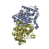 8gqfC 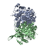 8gqgC 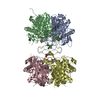 8gqhC 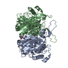 8gqiC 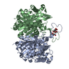 8gqkC  8gqlC  8gqmC 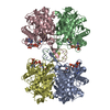 8gqnC 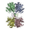 4o99S S: Starting model for refinement C: citing same article ( |
|---|---|
| Similar structure data |
-Links
 PDBj
PDBj

- Assembly
| Deposited unit | 
| ||||||||
|---|---|---|---|---|---|---|---|---|---|
| 1 | 
| ||||||||
| Unit cell |
|
-Components
| #1: Protein | Mass: 43689.730 Da / Num. of mol.: 2 Source method: isolated from a genetically manipulated source Source: (gene. exp.) Pseudomonas aeruginosa PAO1 (bacteria)Strain: ATCC 15692 / DSM 22644 / CIP 104116 / JCM 14847 / LMG 12228 / 1C / PRS 101 / PAO1 Gene: atoB, PA2001 / Production host: Escherichia coli BL21(DE3) (bacteria) / References: UniProt: Q9I2A8, acetyl-CoA C-acetyltransferase#2: Chemical | Glycerol  Mass: 92.094 Da / Num. of mol.: 3 / Source method: obtained synthetically / Formula: C3H8O3 Mass: 92.094 Da / Num. of mol.: 3 / Source method: obtained synthetically / Formula: C3H8O3#3: Chemical | Polyethylene glycol  Mass: 150.173 Da / Num. of mol.: 2 / Source method: obtained synthetically / Formula: C6H14O4 Mass: 150.173 Da / Num. of mol.: 2 / Source method: obtained synthetically / Formula: C6H14O4#4: Chemical | ChemComp-2PE / | Polyethylene glycol  Mass: 414.488 Da / Num. of mol.: 1 / Source method: obtained synthetically / Formula: C18H38O10 / Comment: precipitant*YM Mass: 414.488 Da / Num. of mol.: 1 / Source method: obtained synthetically / Formula: C18H38O10 / Comment: precipitant*YM#5: Water | ChemComp-HOH / | Water Mass: 18.015 Da / Num. of mol.: 643 / Source method: isolated from a natural source / Formula: H2O Mass: 18.015 Da / Num. of mol.: 643 / Source method: isolated from a natural source / Formula: H2OHas ligand of interest | N | |
|---|
-Experimental details
-Experiment
| Experiment | Method: X-RAY DIFFRACTION / Number of used crystals: 1 |
|---|
- Sample preparation
| Crystal | Density Matthews: 3.46 Å3/Da / Density % sol: 64.46 % |
|---|---|
| Crystal grow | Temperature: 293 K / Method: vapor diffusion, hanging drop / Details: 12% (w/v) PEG 3350 4% (v/v) Tacsimate, pH 8.0 |
-Data collection
| Diffraction | Mean temperature: 100 K / Serial crystal experiment: N | |||||||||||||||||||||||||||||||||||||||||||||||||||||||||||||||||||||||||||||||||||||||||||||||||||||||||||||||||||||||||||||||||||||||||||||||||||||||||||||||||||||||||||||||||||||||||||||
|---|---|---|---|---|---|---|---|---|---|---|---|---|---|---|---|---|---|---|---|---|---|---|---|---|---|---|---|---|---|---|---|---|---|---|---|---|---|---|---|---|---|---|---|---|---|---|---|---|---|---|---|---|---|---|---|---|---|---|---|---|---|---|---|---|---|---|---|---|---|---|---|---|---|---|---|---|---|---|---|---|---|---|---|---|---|---|---|---|---|---|---|---|---|---|---|---|---|---|---|---|---|---|---|---|---|---|---|---|---|---|---|---|---|---|---|---|---|---|---|---|---|---|---|---|---|---|---|---|---|---|---|---|---|---|---|---|---|---|---|---|---|---|---|---|---|---|---|---|---|---|---|---|---|---|---|---|---|---|---|---|---|---|---|---|---|---|---|---|---|---|---|---|---|---|---|---|---|---|---|---|---|---|---|---|---|---|---|---|---|---|
| Diffraction source | Source: SYNCHROTRON / Site: PAL/PLS  / Beamline: 7A (6B, 6C1) / Wavelength: 0.9793 Å / Beamline: 7A (6B, 6C1) / Wavelength: 0.9793 Å | |||||||||||||||||||||||||||||||||||||||||||||||||||||||||||||||||||||||||||||||||||||||||||||||||||||||||||||||||||||||||||||||||||||||||||||||||||||||||||||||||||||||||||||||||||||||||||||
| Detector | Type: ADSC QUANTUM 270 / Detector: CCD / Date: Jun 18, 2019 | |||||||||||||||||||||||||||||||||||||||||||||||||||||||||||||||||||||||||||||||||||||||||||||||||||||||||||||||||||||||||||||||||||||||||||||||||||||||||||||||||||||||||||||||||||||||||||||
| Radiation | Protocol: SINGLE WAVELENGTH / Monochromatic (M) / Laue (L): M / Scattering type: x-ray | |||||||||||||||||||||||||||||||||||||||||||||||||||||||||||||||||||||||||||||||||||||||||||||||||||||||||||||||||||||||||||||||||||||||||||||||||||||||||||||||||||||||||||||||||||||||||||||
| Radiation wavelength | Wavelength: 0.9793 Å / Relative weight: 1 | |||||||||||||||||||||||||||||||||||||||||||||||||||||||||||||||||||||||||||||||||||||||||||||||||||||||||||||||||||||||||||||||||||||||||||||||||||||||||||||||||||||||||||||||||||||||||||||
| Reflection | Resolution: 1.78→50 Å / Num. obs: 113500 / % possible obs: 98.1 % / Redundancy: 13 % / Rmerge(I) obs: 0.161 / Rpim(I) all: 0.039 / Rrim(I) all: 0.166 / Χ2: 4.306 / Net I/σ(I): 9.1 | |||||||||||||||||||||||||||||||||||||||||||||||||||||||||||||||||||||||||||||||||||||||||||||||||||||||||||||||||||||||||||||||||||||||||||||||||||||||||||||||||||||||||||||||||||||||||||||
| Reflection shell | Diffraction-ID: 1
|
- Processing
| Software |
| ||||||||||||||||||||||||||||||||||||||||||||||||||||||||||||
|---|---|---|---|---|---|---|---|---|---|---|---|---|---|---|---|---|---|---|---|---|---|---|---|---|---|---|---|---|---|---|---|---|---|---|---|---|---|---|---|---|---|---|---|---|---|---|---|---|---|---|---|---|---|---|---|---|---|---|---|---|---|
| Refinement | Method to determine structure: MOLECULAR REPLACEMENT Starting model: 4O99 Resolution: 1.78→32.75 Å / Cor.coef. Fo:Fc: 0.964 / Cor.coef. Fo:Fc free: 0.954 / SU B: 1.739 / SU ML: 0.052 / Cross valid method: THROUGHOUT / σ(F): 0 / ESU R: 0.079 / ESU R Free: 0.079 / Stereochemistry target values: MAXIMUM LIKELIHOOD Details: HYDROGENS HAVE BEEN ADDED IN THE RIDING POSITIONS U VALUES : REFINED INDIVIDUALLY
| ||||||||||||||||||||||||||||||||||||||||||||||||||||||||||||
| Solvent computation | Ion probe radii: 0.8 Å / Shrinkage radii: 0.8 Å / VDW probe radii: 1.2 Å / Solvent model: MASK | ||||||||||||||||||||||||||||||||||||||||||||||||||||||||||||
| Displacement parameters | Biso max: 96.49 Å2 / Biso mean: 17.436 Å2 / Biso min: 7.87 Å2
| ||||||||||||||||||||||||||||||||||||||||||||||||||||||||||||
| Refinement step | Cycle: final / Resolution: 1.78→32.75 Å
| ||||||||||||||||||||||||||||||||||||||||||||||||||||||||||||
| Refine LS restraints |
| ||||||||||||||||||||||||||||||||||||||||||||||||||||||||||||
| LS refinement shell | Resolution: 1.784→1.831 Å / Rfactor Rfree error: 0 / Total num. of bins used: 20
|
