 Movie
Movie Controller


+ Open data

- Basic information
| Entry | Database: PDB / ID: 8g2f | ||||||
|---|---|---|---|---|---|---|---|
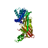
| Title | Crystal Structure of PRMT3 with Compound II710 | ||||||
 Components Components | Protein arginine N-methyltransferase 3 | ||||||
 Keywords Keywords | TRANSFERASE/INHIBITOR /  PRMT3 / II710 / TRANSFERASE / Structural Genomics / PSI-2 / Protein Structure Initiative / Structural Genomics Consortium / SGC / TRANSFERASE-INHIBITOR complex PRMT3 / II710 / TRANSFERASE / Structural Genomics / PSI-2 / Protein Structure Initiative / Structural Genomics Consortium / SGC / TRANSFERASE-INHIBITOR complex | ||||||
| Function / homology |  Function and homology information Function and homology informationprotein-arginine omega-N monomethyltransferase activity / negative regulation of retinoic acid biosynthetic process / type I protein arginine methyltransferase / protein-arginine omega-N asymmetric methyltransferase activity / protein-arginine N-methyltransferase activity / Protein methylation / negative regulation of protein ubiquitination / methyltransferase activity / RMTs methylate histone arginines / ribosome binding ...protein-arginine omega-N monomethyltransferase activity / negative regulation of retinoic acid biosynthetic process / type I protein arginine methyltransferase / protein-arginine omega-N asymmetric methyltransferase activity / protein-arginine N-methyltransferase activity / Protein methylation / negative regulation of protein ubiquitination / methyltransferase activity / RMTs methylate histone arginines / ribosome binding / methylation / metal ion binding / nucleus / cytosol / cytoplasmSimilarity search - Function | ||||||
| Biological species |  Homo sapiens (human) Homo sapiens (human) | ||||||
| Method | X-RAY DIFFRACTION / SYNCHROTRON / MOLECULAR REPLACEMENT / Resolution: 2.06 Å | ||||||
 Authors Authors | Song, X. / Dong, A. / Deng, Y. / Huang, R. / Arrowsmith, C.H. / Edwards, A.M. / Min, J. / Structural Genomics Consortium (SGC) | ||||||
| Funding support |  Canada, 1items Canada, 1items
| ||||||
 Citation Citation | Journal: Acta Pharm Sin B / Year: 2023 Title: A unique binding pocket induced by a noncanonical SAH mimic to develop potent and selective PRMT inhibitors. Authors: Deng, Y. / Song, X. / Iyamu, I.D. / Dong, A. / Min, J. / Huang, R. | ||||||
| History |
|
- Structure visualization
| Structure viewer | Molecule: MolmilJmol/JSmol |
|---|
- Downloads & links
-Download
| PDBx/mmCIF format | 8g2f.cif.gz | 77.4 KB | Display | PDBx/mmCIF format |
|---|---|---|---|---|
| PDB format | pdb8g2f.ent.gz | 54.6 KB | Display | PDB format |
| PDBx/mmJSON format | 8g2f.json.gz | Tree view | PDBx/mmJSON format | |
| Others |  Other downloads Other downloads |
-Validation report
| Arichive directory | https://data.pdbj.org/pub/pdb/validation_reports/g2/8g2fftp://data.pdbj.org/pub/pdb/validation_reports/g2/8g2f | HTTPS FTP |
|---|
-Related structure data
-Links
 PDBj
PDBj


- Assembly
| Deposited unit | 
| ||||||||
|---|---|---|---|---|---|---|---|---|---|
| 1 | 
| ||||||||
| Unit cell |
| ||||||||
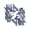
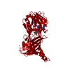
| Details | This construct only comprises the methyltransferase domain of Protein arginine N-methyltransferase 3. The authors do not know the quaternary structure of this domain in isolation. However, the software PISA predicts it to be a dimer. |
-Components
| #1: Protein | Mass: 38281.914 Da / Num. of mol.: 1 Source method: isolated from a genetically manipulated source Source: (gene. exp.) Homo sapiens (human) / Gene: PRMT3, HRMT1L3 / Production host:  Escherichia coli (E. coli) Escherichia coli (E. coli)References: UniProt: O60678, type I protein arginine methyltransferase |
|---|---|
| #2: Chemical | ChemComp-DVS /   Mass: 472.564 Da / Num. of mol.: 1 / Source method: obtained synthetically / Formula: C21H28N8O3S / Feature type: SUBJECT OF INVESTIGATION Mass: 472.564 Da / Num. of mol.: 1 / Source method: obtained synthetically / Formula: C21H28N8O3S / Feature type: SUBJECT OF INVESTIGATION |
| #3: Water | ChemComp-HOH / Water Mass: 18.015 Da / Num. of mol.: 42 / Source method: isolated from a natural source / Formula: H2O Mass: 18.015 Da / Num. of mol.: 42 / Source method: isolated from a natural source / Formula: H2O |
| Has ligand of interest | Y |
-Experimental details
-Experiment
| Experiment | Method: X-RAY DIFFRACTION / Number of used crystals: 1 |
|---|
- Sample preparation
| Crystal | Density Matthews: 2.86 Å3/Da / Density % sol: 57.04 % |
|---|---|
| Crystal grow | Temperature: 291 K / Method: vapor diffusion, hanging drop / Details: 20% PEG3350, 0.3 M Sodium Formate |
-Data collection
| Diffraction | Mean temperature: 100 K / Serial crystal experiment: N |
|---|---|
| Diffraction source | Source: SYNCHROTRON / Site: APS  / Beamline: 24-ID-C / Wavelength: 0.97918 Å / Beamline: 24-ID-C / Wavelength: 0.97918 Å |
| Detector | Type: DECTRIS EIGER2 X 16M / Detector: PIXEL / Date: Mar 16, 2021 |
| Radiation | Protocol: SINGLE WAVELENGTH / Monochromatic (M) / Laue (L): M / Scattering type: x-ray |
| Radiation wavelength | Wavelength: 0.97918 Å / Relative weight: 1 |
| Reflection | Resolution: 2.06→50 Å / Num. obs: 27517 / % possible obs: 97.4 % / Redundancy: 10.4 % / CC1/2: 1 / CC star: 1 / Rmerge(I) obs: 0.09 / Χ2: 1.823 / Net I/av σ(I): 30.9 / Net I/σ(I): 8.9 |
| Reflection shell | Resolution: 2.06→2.1 Å / Redundancy: 8.1 % / Rmerge(I) obs: 0.841 / Mean I/σ(I) obs: 1.7 / Num. unique obs: 1125 / CC1/2: 0.915 / CC star: 0.978 / Χ2: 0.798 / % possible all: 81.6 |
- Processing
| Software |
| ||||||||||||||||||||||||||||||||||||||||||||||||||||||||||||||||||||||||||||||||||||||||||||||||||||||||||||||||||||||||||||||||||||||||||||||||||||||||||||||||||||||||||||||||||||||
|---|---|---|---|---|---|---|---|---|---|---|---|---|---|---|---|---|---|---|---|---|---|---|---|---|---|---|---|---|---|---|---|---|---|---|---|---|---|---|---|---|---|---|---|---|---|---|---|---|---|---|---|---|---|---|---|---|---|---|---|---|---|---|---|---|---|---|---|---|---|---|---|---|---|---|---|---|---|---|---|---|---|---|---|---|---|---|---|---|---|---|---|---|---|---|---|---|---|---|---|---|---|---|---|---|---|---|---|---|---|---|---|---|---|---|---|---|---|---|---|---|---|---|---|---|---|---|---|---|---|---|---|---|---|---|---|---|---|---|---|---|---|---|---|---|---|---|---|---|---|---|---|---|---|---|---|---|---|---|---|---|---|---|---|---|---|---|---|---|---|---|---|---|---|---|---|---|---|---|---|---|---|---|---|
| Refinement | Method to determine structure: MOLECULAR REPLACEMENT / Resolution: 2.06→48.14 Å / Cor.coef. Fo:Fc: 0.965 / Cor.coef. Fo:Fc free: 0.948 / SU B: 5.957 / SU ML: 0.148 / Cross valid method: THROUGHOUT / ESU R: 0.167 / ESU R Free: 0.164 / Stereochemistry target values: MAXIMUM LIKELIHOOD / Details: HYDROGENS HAVE BEEN ADDED IN THE RIDING POSITIONS
| ||||||||||||||||||||||||||||||||||||||||||||||||||||||||||||||||||||||||||||||||||||||||||||||||||||||||||||||||||||||||||||||||||||||||||||||||||||||||||||||||||||||||||||||||||||||
| Solvent computation | Ion probe radii: 0.8 Å / Shrinkage radii: 0.8 Å / VDW probe radii: 1.2 Å / Solvent model: MASK | ||||||||||||||||||||||||||||||||||||||||||||||||||||||||||||||||||||||||||||||||||||||||||||||||||||||||||||||||||||||||||||||||||||||||||||||||||||||||||||||||||||||||||||||||||||||
| Displacement parameters | Biso mean: 54.369 Å2
| ||||||||||||||||||||||||||||||||||||||||||||||||||||||||||||||||||||||||||||||||||||||||||||||||||||||||||||||||||||||||||||||||||||||||||||||||||||||||||||||||||||||||||||||||||||||
| Refinement step | Cycle: 1 / Resolution: 2.06→48.14 Å
| ||||||||||||||||||||||||||||||||||||||||||||||||||||||||||||||||||||||||||||||||||||||||||||||||||||||||||||||||||||||||||||||||||||||||||||||||||||||||||||||||||||||||||||||||||||||
| Refine LS restraints |
|