 Movie
Movie Controller

[English] 日本語
 Yorodumi
- PDB-8fe9: Crystal structure of Ack1 kinase K161Q mutant in complex with the... -
+ Open data

- Basic information
| Entry | Database: PDB / ID: 8fe9 | ||||||||||||
|---|---|---|---|---|---|---|---|---|---|---|---|---|---|
| Title | Crystal structure of Ack1 kinase K161Q mutant in complex with the selective inhibitor (R)-9b | ||||||||||||
 Components Components | Activated CDC42 kinase 1 | ||||||||||||
 Keywords Keywords | TRANSFERASE/TRANSFERASE INHIBITOR /  tyrosine kinase / inhibitor / TRANSFERASE / TRANSFERASE-TRANSFERASE INHIBITOR complex tyrosine kinase / inhibitor / TRANSFERASE / TRANSFERASE-TRANSFERASE INHIBITOR complex | ||||||||||||
| Function / homology |  Function and homology informationregulation of clathrin-dependent endocytosis / cytoophidium / Grb2-EGFR complex / GTPase inhibitor activity / WW domain binding / clathrin-coated vesicle / epidermal growth factor receptor binding / small GTPase-mediated signal transduction / clathrin-coated pit / protein serine/threonine/tyrosine kinase activity ...regulation of clathrin-dependent endocytosis / cytoophidium / Grb2-EGFR complex / GTPase inhibitor activity / WW domain binding / clathrin-coated vesicle / epidermal growth factor receptor binding / small GTPase-mediated signal transduction / clathrin-coated pit / protein serine/threonine/tyrosine kinase activity / adherens junction / non-specific protein-tyrosine kinase / non-membrane spanning protein tyrosine kinase activity / cytoplasmic vesicle membrane / endocytosis / positive regulation of peptidyl-tyrosine phosphorylation / protein tyrosine kinase activity / cell surface receptor signaling pathway / non-specific serine/threonine protein kinase / endosome / phosphorylation / protein serine kinase activity / intracellular membrane-bounded organelle / protein serine/threonine kinase activity / ubiquitin protein ligase binding / perinuclear region of cytoplasm / ATP binding / membrane / identical protein binding / metal ion binding / nucleus / plasma membrane / cytosol / cytoplasm Function and homology informationregulation of clathrin-dependent endocytosis / cytoophidium / Grb2-EGFR complex / GTPase inhibitor activity / WW domain binding / clathrin-coated vesicle / epidermal growth factor receptor binding / small GTPase-mediated signal transduction / clathrin-coated pit / protein serine/threonine/tyrosine kinase activity ...regulation of clathrin-dependent endocytosis / cytoophidium / Grb2-EGFR complex / GTPase inhibitor activity / WW domain binding / clathrin-coated vesicle / epidermal growth factor receptor binding / small GTPase-mediated signal transduction / clathrin-coated pit / protein serine/threonine/tyrosine kinase activity / adherens junction / non-specific protein-tyrosine kinase / non-membrane spanning protein tyrosine kinase activity / cytoplasmic vesicle membrane / endocytosis / positive regulation of peptidyl-tyrosine phosphorylation / protein tyrosine kinase activity / cell surface receptor signaling pathway / non-specific serine/threonine protein kinase / endosome / phosphorylation / protein serine kinase activity / intracellular membrane-bounded organelle / protein serine/threonine kinase activity / ubiquitin protein ligase binding / perinuclear region of cytoplasm / ATP binding / membrane / identical protein binding / metal ion binding / nucleus / plasma membrane / cytosol / cytoplasmSimilarity search - Function | ||||||||||||
| Biological species |  Homo sapiens (human) Homo sapiens (human) | ||||||||||||
| Method | X-RAY DIFFRACTION / SYNCHROTRON / MOLECULAR REPLACEMENT / Resolution: 3.2 Å | ||||||||||||
 Authors Authors | Paung, Y. / Kan, Y. / Seeliger, M.S. / Miller, W.T. | ||||||||||||
| Funding support |  United States, 3items United States, 3items
| ||||||||||||
 Citation Citation | Journal: Biochemistry / Year: 2023 Title: Biochemical Studies of Systemic Lupus Erythematosus-Associated Mutations in Nonreceptor Tyrosine Kinases Ack1 and Brk. Authors: Kan, Y. / Paung, Y. / Kim, Y. / Seeliger, M.A. / Miller, W.T. | ||||||||||||
| History |
|
- Structure visualization
| Structure viewer | Molecule: MolmilJmol/JSmol |
|---|
- Downloads & links
-Download
| PDBx/mmCIF format | 8fe9.cif.gz | 149.2 KB | Display | PDBx/mmCIF format |
|---|---|---|---|---|
| PDB format | pdb8fe9.ent.gz | 101.6 KB | Display | PDB format |
| PDBx/mmJSON format | 8fe9.json.gz | Tree view | PDBx/mmJSON format | |
| Others |  Other downloads Other downloads |
-Validation report
| Arichive directory | https://data.pdbj.org/pub/pdb/validation_reports/fe/8fe9ftp://data.pdbj.org/pub/pdb/validation_reports/fe/8fe9 | HTTPS FTP |
|---|
-Related structure data
| Similar structure data |
|---|
-Links
 PDBj
PDBj
- Assembly
| Deposited unit | 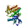
| ||||||||||||
|---|---|---|---|---|---|---|---|---|---|---|---|---|---|
| 1 |
| ||||||||||||
| Unit cell |
|
-Components
| #1: Protein | Mass: 32437.344 Da / Num. of mol.: 1 / Mutation: K161Q Source method: isolated from a genetically manipulated source Source: (gene. exp.) Homo sapiens (human) / Gene: TNK2, ACK1 / Plasmid: pFASTBAC / Cell line (production host): Sf9 / Production host:   Spodoptera frugiperda (fall armyworm) / Tissue (production host): Ovary Spodoptera frugiperda (fall armyworm) / Tissue (production host): OvaryReferences: UniProt: Q07912, non-specific protein-tyrosine kinase, non-specific serine/threonine protein kinase |
|---|---|
| #2: Chemical | ChemComp-R9B / 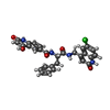  Mass: 542.970 Da / Num. of mol.: 1 / Source method: obtained synthetically / Formula: C29H23ClN4O5 / Feature type: SUBJECT OF INVESTIGATION Mass: 542.970 Da / Num. of mol.: 1 / Source method: obtained synthetically / Formula: C29H23ClN4O5 / Feature type: SUBJECT OF INVESTIGATION |
| #3: Water | ChemComp-HOH / Water Mass: 18.015 Da / Num. of mol.: 5 / Source method: isolated from a natural source / Formula: H2O Mass: 18.015 Da / Num. of mol.: 5 / Source method: isolated from a natural source / Formula: H2O |
| Has ligand of interest | Y |
-Experimental details
-Experiment
| Experiment | Method: X-RAY DIFFRACTION / Number of used crystals: 1 |
|---|
- Sample preparation
| Crystal | Density Matthews: 3.08 Å3/Da / Density % sol: 60.05 % |
|---|---|
| Crystal grow | Temperature: 277 K / Method: vapor diffusion, hanging drop / pH: 6 Details: 0.05 M Bis-Tris (pH 5.8), 19% PEG3350, 0.3 M MgCl2, 2.5% glycerol PH range: 6.0-6.2 |
-Data collection
| Diffraction | Mean temperature: 100 K / Serial crystal experiment: N |
|---|---|
| Diffraction source | Source: SYNCHROTRON / Site: NSLS-II / Beamline: 17-ID-1 / Wavelength: 0.98 Å |
| Detector | Type: DECTRIS EIGER X 9M / Detector: PIXEL / Date: Mar 26, 2021 |
| Radiation | Protocol: SINGLE WAVELENGTH / Monochromatic (M) / Laue (L): M / Scattering type: x-ray |
| Radiation wavelength | Wavelength: 0.98 Å / Relative weight: 1 |
| Reflection | Resolution: 3.2→28.99 Å / Num. obs: 7020 / % possible obs: 99 % / Redundancy: 19.3 % / Rmerge(I) obs: 0.148 / Net I/σ(I): 19.6 |
| Reflection shell | Resolution: 3.2→3.28 Å / Rmerge(I) obs: 1.088 / Num. unique obs: 624 / % possible all: 93 |
- Processing
| Software |
| ||||||||||||||||||||||||||||||||||||||||||
|---|---|---|---|---|---|---|---|---|---|---|---|---|---|---|---|---|---|---|---|---|---|---|---|---|---|---|---|---|---|---|---|---|---|---|---|---|---|---|---|---|---|---|---|
| Refinement | Method to determine structure: MOLECULAR REPLACEMENT / Resolution: 3.2→28.99 Å / SU ML: 0.26 / Cross valid method: FREE R-VALUE / σ(F): 1.34 / Phase error: 30.0648 Stereochemistry target values: GeoStd + Monomer Library + CDL v1.2
| ||||||||||||||||||||||||||||||||||||||||||
| Solvent computation | Shrinkage radii: 0.9 Å / VDW probe radii: 1.11 Å / Solvent model: FLAT BULK SOLVENT MODEL | ||||||||||||||||||||||||||||||||||||||||||
| Refinement step | Cycle: LAST / Resolution: 3.2→28.99 Å
| ||||||||||||||||||||||||||||||||||||||||||
| Refine LS restraints |
| ||||||||||||||||||||||||||||||||||||||||||
| LS refinement shell |
| ||||||||||||||||||||||||||||||||||||||||||
| Refinement TLS params. | Method: refined / Origin x: 33.8745121239 Å / Origin y: 18.4514654772 Å / Origin z: 24.6697009095 Å
| ||||||||||||||||||||||||||||||||||||||||||
| Refinement TLS group | Selection details: all |
