 Movie
Movie Controller


+ Open data

- Basic information
| Entry | Database: PDB / ID: 8f47 | ||||||
|---|---|---|---|---|---|---|---|
| Title | Crystal structure of VACV D13 in complex with STK69439 | ||||||
 Components Components | Scaffold protein D13 Scaffolding Scaffolding | ||||||
 Keywords Keywords | VIRAL PROTEIN / poxvirus / assembly / scaffolding protein / Fragment-based drug design / immature virion | ||||||
| Function / homology | Poxvirus rifampicin-resistance / Poxvirus rifampicin resistance protein / response to antibiotic / membrane / identical protein binding / FORMIC ACID / 6,8-dimethoxy-2-methylquinolin-4-amine / Scaffold protein OPG125 Function and homology information Function and homology information | ||||||
| Biological species |   Vaccinia virus Vaccinia virus | ||||||
| Method | X-RAY DIFFRACTION / SYNCHROTRON / MOLECULAR REPLACEMENT / Resolution: 3.1 Å | ||||||
 Authors Authors | Subedi, B.P. / Garriga, D. / Coulibaly, F. | ||||||
| Funding support |  Australia, 1items Australia, 1items
| ||||||
 Citation Citation | Journal: To Be Published Title: Structure of scaffolidng protein D13 of Vaccinia Virus in complex with fragments inhibiting A17 binding. Authors: Subedi, B.P. / Garriga, D. / Coulibaly, F. #1: Journal: Proc Natl Acad Sci U S A / Year: 2018Title: Structural basis for the inhibition of poxvirus assembly by the antibiotic rifampicin. Authors: Garriga, D. / Headey, S. / Accurso, C. / Gunzburg, M. / Scanlon, M. / Coulibaly, F. | ||||||
| History |
|
- Structure visualization
| Structure viewer | Molecule: MolmilJmol/JSmol |
|---|
- Downloads & links
-Download
| PDBx/mmCIF format | 8f47.cif.gz | 411.4 KB | Display | PDBx/mmCIF format |
|---|---|---|---|---|
| PDB format | pdb8f47.ent.gz | 270 KB | Display | PDB format |
| PDBx/mmJSON format | 8f47.json.gz | Tree view | PDBx/mmJSON format | |
| Others |  Other downloads Other downloads |
-Validation report
| Arichive directory | https://data.pdbj.org/pub/pdb/validation_reports/f4/8f47ftp://data.pdbj.org/pub/pdb/validation_reports/f4/8f47 | HTTPS FTP |
|---|
-Related structure data
| Related structure data |  8f65C 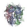 6beiS S: Starting model for refinement C: citing same article ( |
|---|---|
| Similar structure data |
-Links
 PDBj
PDBj- Assembly
| Deposited unit | 
| ||||||||||||||||||||||||||||||||||||||||||||||||||||||||||||||||||||||||||||||||||||||||||||||||||||||||||||||||||
|---|---|---|---|---|---|---|---|---|---|---|---|---|---|---|---|---|---|---|---|---|---|---|---|---|---|---|---|---|---|---|---|---|---|---|---|---|---|---|---|---|---|---|---|---|---|---|---|---|---|---|---|---|---|---|---|---|---|---|---|---|---|---|---|---|---|---|---|---|---|---|---|---|---|---|---|---|---|---|---|---|---|---|---|---|---|---|---|---|---|---|---|---|---|---|---|---|---|---|---|---|---|---|---|---|---|---|---|---|---|---|---|---|---|---|---|
| 1 |
| ||||||||||||||||||||||||||||||||||||||||||||||||||||||||||||||||||||||||||||||||||||||||||||||||||||||||||||||||||
| Unit cell |
| ||||||||||||||||||||||||||||||||||||||||||||||||||||||||||||||||||||||||||||||||||||||||||||||||||||||||||||||||||
| Noncrystallographic symmetry (NCS) | NCS domain:
NCS domain segments:
|