 Movie
Movie Controller

[English] 日本語
 Yorodumi
- PDB-8evi: CX3CR1 nucleosome and PU.1 complex containing disulfide bond mutations -
+ Open data

- Basic information
| Entry | Database: PDB / ID: 8evi | ||||||
|---|---|---|---|---|---|---|---|
| Title | CX3CR1 nucleosome and PU.1 complex containing disulfide bond mutations | ||||||
 Components Components |
| ||||||
 Keywords Keywords | TRANSCRIPTION/DNA /  nucleosome / transcription factor / transcription / chromatin binding protein-DNA complex / TRANSCRIPTION-DNA complex nucleosome / transcription factor / transcription / chromatin binding protein-DNA complex / TRANSCRIPTION-DNA complex | ||||||
| Function / homology |  Function and homology information Function and homology informationpositive regulation of myeloid dendritic cell chemotaxis / anatomical structure regression / follicular B cell differentiation / positive regulation of antifungal innate immune response / regulation of myeloid progenitor cell differentiation / pro-T cell differentiation / negative regulation of neutrophil degranulation / germinal center B cell differentiation / myeloid leukocyte differentiation / positive regulation of microglial cell mediated cytotoxicity ...positive regulation of myeloid dendritic cell chemotaxis / anatomical structure regression / follicular B cell differentiation / positive regulation of antifungal innate immune response / regulation of myeloid progenitor cell differentiation / pro-T cell differentiation / negative regulation of neutrophil degranulation / germinal center B cell differentiation / myeloid leukocyte differentiation / positive regulation of microglial cell mediated cytotoxicity / granulocyte differentiation / lymphocyte differentiation / apoptotic process involved in blood vessel morphogenesis / endothelial to hematopoietic transition / negative regulation of adipose tissue development / pericyte cell differentiation / immature B cell differentiation / negative regulation of MHC class II biosynthetic process / lymphoid progenitor cell differentiation / myeloid dendritic cell differentiation / vasculature development / regulation of DNA-binding transcription factor activity / oncogene-induced cell senescence / negative regulation of protein localization to chromatin / positive regulation of p38MAPK cascade / positive regulation of B cell differentiation / NFAT protein binding / somatic stem cell population maintenance / macrophage differentiation / negative regulation of megakaryocyte differentiation / protein localization to CENP-A containing chromatin / cis-regulatory region sequence-specific DNA binding / Chromatin modifying enzymes / Replacement of protamines by nucleosomes in the male pronucleus / CENP-A containing nucleosome / epigenetic regulation of gene expression / Packaging Of Telomere Ends / Recognition and association of DNA glycosylase with site containing an affected purine / Cleavage of the damaged purine / Deposition of new CENPA-containing nucleosomes at the centromere / lipopolysaccharide-mediated signaling pathway / transcription initiation-coupled chromatin remodeling / Recognition and association of DNA glycosylase with site containing an affected pyrimidine / Cleavage of the damaged pyrimidine / Inhibition of DNA recombination at telomere / Meiotic synapsis / telomere organization / protein sequestering activity / RNA Polymerase I Promoter Opening / Interleukin-7 signaling / Assembly of the ORC complex at the origin of replication / SUMOylation of chromatin organization proteins / erythrocyte differentiation / transforming growth factor beta receptor signaling pathway / DNA methylation / Condensation of Prophase Chromosomes / ERCC6 (CSB) and EHMT2 (G9a) positively regulate rRNA expression / SIRT1 negatively regulates rRNA expression / Chromatin modifications during the maternal to zygotic transition (MZT) / HCMV Late Events / innate immune response in mucosa / PRC2 methylates histones and DNA / Defective pyroptosis / HDACs deacetylate histones / RNA Polymerase I Promoter Escape / Nonhomologous End-Joining (NHEJ) / Transcriptional regulation by small RNAs / Formation of the beta-catenin:TCF transactivating complex / RUNX1 regulates genes involved in megakaryocyte differentiation and platelet function / Activated PKN1 stimulates transcription of AR (androgen receptor) regulated genes KLK2 and KLK3 / NoRC negatively regulates rRNA expression / G2/M DNA damage checkpoint / B-WICH complex positively regulates rRNA expression / HDMs demethylate histones / DNA Damage/Telomere Stress Induced Senescence / Metalloprotease DUBs / PKMTs methylate histone lysines / RMTs methylate histone arginines / Meiotic recombination / Pre-NOTCH Transcription and Translation / DNA-binding transcription repressor activity, RNA polymerase II-specific / histone deacetylase binding / positive regulation of miRNA transcription / Activation of anterior HOX genes in hindbrain development during early embryogenesis / HCMV Early Events / Transcriptional regulation of granulopoiesis / structural constituent of chromatin / UCH proteinases / nucleosome / antimicrobial humoral immune response mediated by antimicrobial peptide / nucleosome assembly / Recruitment and ATM-mediated phosphorylation of repair and signaling proteins at DNA double strand breaks / RUNX1 regulates transcription of genes involved in differentiation of HSCs / chromatin organization / Factors involved in megakaryocyte development and platelet production / HATs acetylate histones / gene expression / Processing of DNA double-strand break ends / Senescence-Associated Secretory Phenotype (SASP) / antibacterial humoral responseSimilarity search - Function | ||||||
| Biological species |  Homo sapiens (human) Homo sapiens (human) Escherichia coli (E. coli) Escherichia coli (E. coli) Mus musculus (house mouse) Mus musculus (house mouse) | ||||||
| Method | ELECTRON MICROSCOPY / single particle reconstruction / cryo EM / Resolution: 2.64 Å | ||||||
 Authors Authors | Lian, T. / Guan, R. / Bai, Y. | ||||||
| Funding support |  United States, 1items United States, 1items
| ||||||
 Citation Citation | Journal: Nat Struct Mol Biol / Year: 2024 Title: Structural mechanism of synergistic targeting of the CX3CR1 nucleosome by PU.1 and C/EBPα. Authors: Tengfei Lian / Ruifang Guan / Bing-Rui Zhou / Yawen Bai / Abstract: Pioneer transcription factors are vital for cell fate changes. PU.1 and C/EBPα work together to regulate hematopoietic stem cell differentiation. However, how they recognize in vivo nucleosomal DNA ...Pioneer transcription factors are vital for cell fate changes. PU.1 and C/EBPα work together to regulate hematopoietic stem cell differentiation. However, how they recognize in vivo nucleosomal DNA targets remains elusive. Here we report the structures of the nucleosome containing the mouse genomic CX3CR1 enhancer DNA and its complexes with PU.1 alone and with both PU.1 and the C/EBPα DNA binding domain. Our structures reveal that PU.1 binds the DNA motif at the exit linker, shifting 17 bp of DNA into the core region through interactions with H2A, unwrapping ~20 bp of nucleosomal DNA. C/EBPα binding, aided by PU.1's repositioning, unwraps ~25 bp of entry DNA. The PU.1 Q218H mutation, linked to acute myeloid leukemia, disrupts PU.1-H2A interactions. PU.1 and C/EBPα jointly displace linker histone H1 and open the H1-condensed nucleosome array. Our study unveils how two pioneer factors can work cooperatively to open closed chromatin by altering DNA positioning in the nucleosome. | ||||||
| History |
|
- Structure visualization
| Structure viewer | Molecule: MolmilJmol/JSmol |
|---|
- Downloads & links
-Download
| PDBx/mmCIF format | 8evi.cif.gz | 420.3 KB | Display | PDBx/mmCIF format |
|---|---|---|---|---|
| PDB format | pdb8evi.ent.gz | 321.5 KB | Display | PDB format |
| PDBx/mmJSON format | 8evi.json.gz | Tree view | PDBx/mmJSON format | |
| Others |  Other downloads Other downloads |
-Validation report
| Arichive directory | https://data.pdbj.org/pub/pdb/validation_reports/ev/8eviftp://data.pdbj.org/pub/pdb/validation_reports/ev/8evi | HTTPS FTP |
|---|
-Related structure data
| Related structure data | 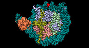 28630MC  8evhC 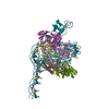 8evjC  8sypC M: map data used to model this data C: citing same article ( |
|---|---|
| Similar structure data |

-Links
 PDBj
PDBj


- Assembly
| Deposited unit | 
|
|---|---|
| 1 |
|
-Components
-DNA chain , 2 types, 2 molecules JI
| #1: DNA chain | Mass: 51538.973 Da / Num. of mol.: 1 / Source method: obtained synthetically / Source: (synth.) Mus musculus (house mouse) |
|---|---|
| #2: DNA chain | Mass: 51558.816 Da / Num. of mol.: 1 / Source method: obtained synthetically / Source: (synth.) Mus musculus (house mouse) |
-Protein , 4 types, 7 molecules AEBFDHO
| #3: Protein | Histone H3 / Histone H3/a / Histone H3/b / Histone H3/c / Histone H3/d / Histone H3/f / Histone H3/h / Histone ...Histone H3/a / Histone H3/b / Histone H3/c / Histone H3/d / Histone H3/f / Histone H3/h / Histone H3/i / Histone H3/j / Histone H3/k / Histone H3/l Mass: 15437.167 Da / Num. of mol.: 2 Source method: isolated from a genetically manipulated source Source: (gene. exp.) Homo sapiens (human)Gene: H3C1, H3FA, HIST1H3A, H3C2, H3FL, HIST1H3B, H3C3, H3FC HIST1H3C, H3C4, H3FB, HIST1H3D, H3C6, H3FD, HIST1H3E, H3C7, H3FI, HIST1H3F, H3C8, H3FH, HIST1H3G, H3C10, H3FK, HIST1H3H, H3C11, H3FF, ...Gene: H3C1, H3FA, HIST1H3A, H3C2, H3FL, HIST1H3B, H3C3, H3FC HIST1H3C, H3C4, H3FB, HIST1H3D, H3C6, H3FD, HIST1H3E, H3C7, H3FI, HIST1H3F, H3C8, H3FH, HIST1H3G, H3C10, H3FK, HIST1H3H, H3C11, H3FF, HIST1H3I, H3C12, H3FJ, HIST1H3J Production host: Escherichia coli 'BL21-Gold(DE3)pLysS AG' (bacteria)References: UniProt: P68431 #4: Protein | Mass: 11394.426 Da / Num. of mol.: 2 Source method: isolated from a genetically manipulated source Source: (gene. exp.) Homo sapiens (human)Gene: HIST1H4A, H4/A, H4FA, HIST1H4B, H4/I, H4FI, HIST1H4C, H4/G, H4FG, HIST1H4D, H4/B, H4FB, HIST1H4E, H4/J, H4FJ, HIST1H4F, H4/C, H4FC, HIST1H4H, H4/H, H4FH, HIST1H4I, H4/M, H4FM, HIST1H4J, H4/E, ...Gene: HIST1H4A, H4/A, H4FA, HIST1H4B, H4/I, H4FI, HIST1H4C, H4/G, H4FG, HIST1H4D, H4/B, H4FB, HIST1H4E, H4/J, H4FJ, HIST1H4F, H4/C, H4FC, HIST1H4H, H4/H, H4FH, HIST1H4I, H4/M, H4FM, HIST1H4J, H4/E, H4FE, HIST1H4K, H4/D, H4FD, HIST1H4L, H4/K, H4FK, HIST2H4A, H4/N, H4F2, H4FN, HIST2H4, HIST2H4B, H4/O, H4FO, HIST4H4 Production host: Escherichia coli 'BL21-Gold(DE3)pLysS AG' (bacteria)References: UniProt: P62805 #6: Protein | Mass: 13951.239 Da / Num. of mol.: 2 Source method: isolated from a genetically manipulated source Source: (gene. exp.) Homo sapiens (human) / Gene: H2BC21, H2BFQ, HIST2H2BEProduction host: Escherichia coli 'BL21-Gold(DE3)pLysS AG' (bacteria)References: UniProt: Q16778 #9: Protein | | Mass: 32865.844 Da / Num. of mol.: 1 / Mutation: G220C Source method: isolated from a genetically manipulated source Source: (gene. exp.) Mus musculus (house mouse) / Gene: Spi1, Sfpi-1, Sfpi1Production host: Escherichia coli 'BL21-Gold(DE3)pLysS AG' (bacteria)References: UniProt: P17433 |
|---|
-Histone H2A type 2- ... , 2 types, 2 molecules CG
| #5: Protein | Mass: 14019.467 Da / Num. of mol.: 1 / Mutation: T77C Source method: isolated from a genetically manipulated source Source: (gene. exp.) Homo sapiens (human) / Gene: H2AC20, H2AFQ, HIST2H2ACProduction host: Escherichia coli 'BL21-Gold(DE3)pLysS AG' (bacteria)References: UniProt: Q16777 |
|---|---|
| #7: Protein | Mass: 14017.428 Da / Num. of mol.: 1 Source method: isolated from a genetically manipulated source Source: (gene. exp.) Homo sapiens (human) / Gene: H2AC20, H2AFQ, HIST2H2ACProduction host: Escherichia coli 'BL21-Gold(DE3)pLysS AG' (bacteria)References: UniProt: Q16777 |
-Antibody , 1 types, 2 molecules MN
| #8: Antibody | Mass: 29030.146 Da / Num. of mol.: 2 Source method: isolated from a genetically manipulated source Source: (gene. exp.) Escherichia coli (E. coli)Production host: Escherichia coli 'BL21-Gold(DE3)pLysS AG' (bacteria) |
|---|
-Experimental details
-Experiment
| Experiment | Method: ELECTRON MICROSCOPY |
|---|---|
| EM experiment | Aggregation state: PARTICLE / 3D reconstruction method: single particle reconstruction |
- Sample preparation
| Component | Name: nucleosome PU.1 mutant complex / Type: COMPLEX / Entity ID: all / Source: MULTIPLE SOURCES | ||||||||||||||||
|---|---|---|---|---|---|---|---|---|---|---|---|---|---|---|---|---|---|
| Source (natural) |
| ||||||||||||||||
| Source (recombinant) | Organism: Escherichia coli 'BL21-Gold(DE3)pLysS AG' (bacteria) | ||||||||||||||||
| Buffer solution | pH: 7.3 | ||||||||||||||||
| Specimen | Embedding applied: NO / Shadowing applied: NO / Staining applied: NO / Vitrification applied: YES | ||||||||||||||||
| Vitrification | Cryogen name: ETHANE |
- Electron microscopy imaging
| Experimental equipment |  Model: Titan Krios / Image courtesy: FEI Company |
|---|---|
| Microscopy | Model: FEI TITAN KRIOS |
| Electron gun | Electron source: FIELD EMISSION GUN / Accelerating voltage: 300 kV / Illumination mode: SPOT SCAN |
| Electron lens | Mode: BRIGHT FIELDBright-field microscopy / Nominal defocus max: 2000 nm / Nominal defocus min: 1000 nm |
| Image recording | Electron dose: 53.8 e/Å2 / Film or detector model: GATAN K3 (6k x 4k) |
- Processing
| CTF correction | Type: NONE |
|---|---|
| 3D reconstruction | Resolution: 2.64 Å / Resolution method: FSC 0.143 CUT-OFF / Num. of particles: 127327 / Symmetry type: POINT |
