 Movie
Movie Controller


+ Open data

- Basic information
| Entry | Database: PDB / ID: 8ese | ||||||
|---|---|---|---|---|---|---|---|
| Title | Crystal structure of human Vps29 bound to a peptide from Vps35L | ||||||
 Components Components |
| ||||||
 Keywords Keywords |  ENDOCYTOSIS / Commander / Retriever / Vps29 / Vps35L ENDOCYTOSIS / Commander / Retriever / Vps29 / Vps35L | ||||||
| Function / homology |  Function and homology informationretromer, cargo-selective complex / WNT ligand biogenesis and trafficking / retromer complex / endocytic recycling / retrograde transport, endosome to Golgi / intracellular protein transport / late endosome / protein transport / early endosome / endosome membrane ...retromer, cargo-selective complex / WNT ligand biogenesis and trafficking / retromer complex / endocytic recycling / retrograde transport, endosome to Golgi / intracellular protein transport / late endosome / protein transport / early endosome / endosome membrane / endosome / intracellular membrane-bounded organelle / metal ion binding / cytosol Function and homology informationretromer, cargo-selective complex / WNT ligand biogenesis and trafficking / retromer complex / endocytic recycling / retrograde transport, endosome to Golgi / intracellular protein transport / late endosome / protein transport / early endosome / endosome membrane ...retromer, cargo-selective complex / WNT ligand biogenesis and trafficking / retromer complex / endocytic recycling / retrograde transport, endosome to Golgi / intracellular protein transport / late endosome / protein transport / early endosome / endosome membrane / endosome / intracellular membrane-bounded organelle / metal ion binding / cytosolSimilarity search - Function | ||||||
| Biological species |  Homo sapiens (human) Homo sapiens (human) | ||||||
| Method | X-RAY DIFFRACTION / SYNCHROTRON / MOLECULAR REPLACEMENT / Resolution: 1.35 Å | ||||||
 Authors Authors | Healy, M.D. / Collins, B.M. | ||||||
| Funding support | 1items
| ||||||
 Citation Citation | Journal: Cell / Year: 2023 Title: Structure of the endosomal Commander complex linked to Ritscher-Schinzel syndrome. Authors: Michael D Healy / Kerrie E McNally / Rebeka Butkovič / Molly Chilton / Kohji Kato / Joanna Sacharz / Calum McConville / Edmund R R Moody / Shrestha Shaw / Vicente J Planelles-Herrero / ...Authors: Michael D Healy / Kerrie E McNally / Rebeka Butkovič / Molly Chilton / Kohji Kato / Joanna Sacharz / Calum McConville / Edmund R R Moody / Shrestha Shaw / Vicente J Planelles-Herrero / Sathish K N Yadav / Jennifer Ross / Ufuk Borucu / Catherine S Palmer / Kai-En Chen / Tristan I Croll / Ryan J Hall / Nikeisha J Caruana / Rajesh Ghai / Thi H D Nguyen / Kate J Heesom / Shinji Saitoh / Imre Berger / Christiane Schaffitzel / Tom A Williams / David A Stroud / Emmanuel Derivery / Brett M Collins / Peter J Cullen /    Abstract: The Commander complex is required for endosomal recycling of diverse transmembrane cargos and is mutated in Ritscher-Schinzel syndrome. It comprises two sub-assemblies: Retriever composed of VPS35L, ...The Commander complex is required for endosomal recycling of diverse transmembrane cargos and is mutated in Ritscher-Schinzel syndrome. It comprises two sub-assemblies: Retriever composed of VPS35L, VPS26C, and VPS29; and the CCC complex which contains twelve subunits: COMMD1-COMMD10 and the coiled-coil domain-containing (CCDC) proteins CCDC22 and CCDC93. Combining X-ray crystallography, electron cryomicroscopy, and in silico predictions, we have assembled a complete structural model of Commander. Retriever is distantly related to the endosomal Retromer complex but has unique features preventing the shared VPS29 subunit from interacting with Retromer-associated factors. The COMMD proteins form a distinctive hetero-decameric ring stabilized by extensive interactions with CCDC22 and CCDC93. These adopt a coiled-coil structure that connects the CCC and Retriever assemblies and recruits a 16th subunit, DENND10, to form the complete Commander complex. The structure allows mapping of disease-causing mutations and reveals the molecular features required for the function of this evolutionarily conserved trafficking machinery. | ||||||
| History |
|
- Structure visualization
| Structure viewer | Molecule: MolmilJmol/JSmol |
|---|
- Downloads & links
-Download
| PDBx/mmCIF format | 8ese.cif.gz | 117.8 KB | Display | PDBx/mmCIF format |
|---|---|---|---|---|
| PDB format | pdb8ese.ent.gz | 73.7 KB | Display | PDB format |
| PDBx/mmJSON format | 8ese.json.gz | Tree view | PDBx/mmJSON format | |
| Others |  Other downloads Other downloads |
-Validation report
| Arichive directory | https://data.pdbj.org/pub/pdb/validation_reports/es/8eseftp://data.pdbj.org/pub/pdb/validation_reports/es/8ese | HTTPS FTP |
|---|
-Related structure data
| Related structure data |  8esdC  8f2rC  8f2uC  6xsaS S: Starting model for refinement C: citing same article ( |
|---|---|
| Similar structure data |


-Links
 PDBj
PDBj
- Assembly
| Deposited unit | 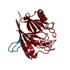
| ||||||||||||
|---|---|---|---|---|---|---|---|---|---|---|---|---|---|
| 1 |
| ||||||||||||
| Unit cell |
|
-Components
| #1: Protein/peptide | Mass: 2635.021 Da / Num. of mol.: 1 Source method: isolated from a genetically manipulated source Source: (gene. exp.) Homo sapiens (human) / Production host:  Escherichia coli (E. coli) / References: UniProt: B3KT69 Escherichia coli (E. coli) / References: UniProt: B3KT69 |
|---|---|
| #2: Protein | Vacuole / hVPS29 / PEP11 homolog / Vesicle protein sorting 29 Mass: 20726.904 Da / Num. of mol.: 1 Source method: isolated from a genetically manipulated source Source: (gene. exp.) Homo sapiens (human) / Gene: VPS29, DC15, DC7, MDS007 / Production host: Escherichia coli (E. coli) / References: UniProt: Q9UBQ0 |
| #3: Water | ChemComp-HOH / Water Mass: 18.015 Da / Num. of mol.: 191 / Source method: isolated from a natural source / Formula: H2O Mass: 18.015 Da / Num. of mol.: 191 / Source method: isolated from a natural source / Formula: H2O |
-Experimental details
-Experiment
| Experiment | Method: X-RAY DIFFRACTION / Number of used crystals: 1 |
|---|
- Sample preparation
| Crystal | Density Matthews: 2.14 Å3/Da / Density % sol: 42.46 % |
|---|---|
| Crystal grow | Temperature: 293 K / Method: vapor diffusion, hanging drop / pH: 5.5 / Details: 0.1 M Bis-tris pH 5.5 and 25% (w/v) PEG3350 |
-Data collection
| Diffraction | Mean temperature: 100 K / Serial crystal experiment: N |
|---|---|
| Diffraction source | Source: SYNCHROTRON / Site: Australian Synchrotron / Beamline: MX2 / Wavelength: 0.95365 Å |
| Detector | Type: DECTRIS EIGER2 X 16M / Detector: PIXEL / Date: Oct 13, 2022 |
| Radiation | Protocol: SINGLE WAVELENGTH / Monochromatic (M) / Laue (L): M / Scattering type: x-ray |
| Radiation wavelength | Wavelength: 0.95365 Å / Relative weight: 1 |
| Reflection | Resolution: 1.35→45.95 Å / Num. obs: 44459 / % possible obs: 99.1 % / Redundancy: 6.2 % / Biso Wilson estimate: 18.43 Å2 / CC1/2: 0.998 / Rpim(I) all: 0.031 / Net I/σ(I): 11.3 |
| Reflection shell | Resolution: 1.35→1.37 Å / Num. unique obs: 1992 / CC1/2: 0.481 / Rpim(I) all: 0.643 |
- Processing
| Software |
| |||||||||||||||||||||||||||||||||||||||||||||||||||||||||||||||||||||||||||||||||||||||||||||||||||||||||
|---|---|---|---|---|---|---|---|---|---|---|---|---|---|---|---|---|---|---|---|---|---|---|---|---|---|---|---|---|---|---|---|---|---|---|---|---|---|---|---|---|---|---|---|---|---|---|---|---|---|---|---|---|---|---|---|---|---|---|---|---|---|---|---|---|---|---|---|---|---|---|---|---|---|---|---|---|---|---|---|---|---|---|---|---|---|---|---|---|---|---|---|---|---|---|---|---|---|---|---|---|---|---|---|---|---|---|
| Refinement | Method to determine structure: MOLECULAR REPLACEMENT Starting model: 6XSA Resolution: 1.35→45.95 Å / SU ML: 0.2316 / Cross valid method: FREE R-VALUE / σ(F): 1.33 / Phase error: 27.422 Stereochemistry target values: GeoStd + Monomer Library + CDL v1.2
| |||||||||||||||||||||||||||||||||||||||||||||||||||||||||||||||||||||||||||||||||||||||||||||||||||||||||
| Solvent computation | Shrinkage radii: 0.9 Å / VDW probe radii: 1.11 Å / Solvent model: FLAT BULK SOLVENT MODEL | |||||||||||||||||||||||||||||||||||||||||||||||||||||||||||||||||||||||||||||||||||||||||||||||||||||||||
| Displacement parameters | Biso mean: 26.67 Å2 | |||||||||||||||||||||||||||||||||||||||||||||||||||||||||||||||||||||||||||||||||||||||||||||||||||||||||
| Refinement step | Cycle: LAST / Resolution: 1.35→45.95 Å
| |||||||||||||||||||||||||||||||||||||||||||||||||||||||||||||||||||||||||||||||||||||||||||||||||||||||||
| Refine LS restraints |
| |||||||||||||||||||||||||||||||||||||||||||||||||||||||||||||||||||||||||||||||||||||||||||||||||||||||||
| LS refinement shell |
|
