 Movie
Movie Controller


+ Open data

- Basic information
| Entry | Database: PDB / ID: 8esa | ||||||
|---|---|---|---|---|---|---|---|
| Title | CryoEM structure of HLA-A2 bound to MAGEA4 (230-239) peptide | ||||||
 Components Components | Beta-2-microglobulin,HLA class I antigen,MAGE-A4 peptide chimera | ||||||
 Keywords Keywords |  IMMUNE SYSTEM / HLA / MHC IMMUNE SYSTEM / HLA / MHC | ||||||
| Function / homology |  Function and homology information Function and homology informationantigen processing and presentation / lumenal side of endoplasmic reticulum membrane / ER to Golgi transport vesicle membrane / recycling endosome membrane / phagocytic vesicle membrane / early endosome membrane / immune response / cell surfaceSimilarity search - Function | ||||||
| Biological species |  Homo sapiens (human) Homo sapiens (human) | ||||||
| Method | ELECTRON MICROSCOPY / single particle reconstruction / cryo EM / Resolution: 3.4 Å | ||||||
 Authors Authors | Saotome, K. / Franklin, M.C. | ||||||
| Funding support | 1items
| ||||||
 Citation Citation | Journal: Nat Commun / Year: 2023 Title: Structural analysis of cancer-relevant TCR-CD3 and peptide-MHC complexes by cryoEM. Authors: Kei Saotome / Drew Dudgeon / Kiersten Colotti / Michael J Moore / Jennifer Jones / Yi Zhou / Ashique Rafique / George D Yancopoulos / Andrew J Murphy / John C Lin / William C Olson / Matthew C Franklin /  Abstract: The recognition of antigenic peptide-MHC (pMHC) molecules by T-cell receptors (TCR) initiates the T-cell mediated immune response. Structural characterization is key for understanding the specificity ...The recognition of antigenic peptide-MHC (pMHC) molecules by T-cell receptors (TCR) initiates the T-cell mediated immune response. Structural characterization is key for understanding the specificity of TCR-pMHC interactions and informing the development of therapeutics. Despite the rapid rise of single particle cryoelectron microscopy (cryoEM), x-ray crystallography has remained the preferred method for structure determination of TCR-pMHC complexes. Here, we report cryoEM structures of two distinct full-length α/β TCR-CD3 complexes bound to their pMHC ligand, the cancer-testis antigen HLA-A2/MAGEA4 (230-239). We also determined cryoEM structures of pMHCs containing MAGEA4 (230-239) peptide and the closely related MAGEA8 (232-241) peptide in the absence of TCR, which provided a structural explanation for the MAGEA4 preference displayed by the TCRs. These findings provide insights into the TCR recognition of a clinically relevant cancer antigen and demonstrate the utility of cryoEM for high-resolution structural analysis of TCR-pMHC interactions. | ||||||
| History |
|
- Structure visualization
| Structure viewer | Molecule: MolmilJmol/JSmol |
|---|
- Downloads & links
-Download
| PDBx/mmCIF format | 8esa.cif.gz | 100 KB | Display | PDBx/mmCIF format |
|---|---|---|---|---|
| PDB format | pdb8esa.ent.gz | 64.4 KB | Display | PDB format |
| PDBx/mmJSON format | 8esa.json.gz | Tree view | PDBx/mmJSON format | |
| Others |  Other downloads Other downloads |
-Validation report
| Arichive directory | https://data.pdbj.org/pub/pdb/validation_reports/es/8esaftp://data.pdbj.org/pub/pdb/validation_reports/es/8esa | HTTPS FTP |
|---|
-Related structure data
| Related structure data |  28573MC  8es7C  8es8C 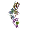 8es9C 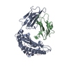 8esbC M: map data used to model this data C: citing same article ( |
|---|---|
| Similar structure data |


-Links
 PDBj
PDBj

- Assembly
| Deposited unit | 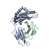
|
|---|---|
| 1 |
|
-Components
| #1: Protein | Mass: 50306.184 Da / Num. of mol.: 3 Source method: isolated from a genetically manipulated source Source: (gene. exp.) Homo sapiens (human) / Gene: HLA-A, HLA / Production host:   Cricetulus griseus (Chinese hamster) / References: UniProt: Q53Z42 Cricetulus griseus (Chinese hamster) / References: UniProt: Q53Z42 |
|---|
-Experimental details
-Experiment
| Experiment | Method: ELECTRON MICROSCOPY |
|---|---|
| EM experiment | Aggregation state: PARTICLE / 3D reconstruction method: single particle reconstruction |
- Sample preparation
| Component | Name: Single chain disulfide stabilized trimer of HLA-A2, Beta-2-microglobulin, MAGE-A4 peptide in complex with anti-Beta-2-microglobulin Fab 2M2 Type: COMPLEX / Entity ID: all / Source: RECOMBINANT |
|---|---|
| Molecular weight | Experimental value: NO |
| Source (natural) | Organism: Homo sapiens (human) |
| Source (recombinant) | Organism: Cricetulus griseus (Chinese hamster) |
| Buffer solution | pH: 7.5 |
| Specimen | Embedding applied: NO / Shadowing applied: NO / Staining applied: NO / Vitrification applied: YES |
| Vitrification | Cryogen name: ETHANE |
- Electron microscopy imaging
| Experimental equipment |  Model: Titan Krios / Image courtesy: FEI Company |
|---|---|
| Microscopy | Model: FEI TITAN KRIOS |
| Electron gun | Electron source: FIELD EMISSION GUN / Accelerating voltage: 300 kV / Illumination mode: FLOOD BEAM |
| Electron lens | Mode: BRIGHT FIELDBright-field microscopy / Nominal defocus max: 2400 nm / Nominal defocus min: 1400 nm |
| Image recording | Electron dose: 40 e/Å2 / Film or detector model: GATAN K3 BIOQUANTUM (6k x 4k) |
| EM imaging optics | Energyfilter name: GIF Bioquantum / Energyfilter slit width: 20 eV |
- Processing
| Software |
| ||||||||||||||||||||||||
|---|---|---|---|---|---|---|---|---|---|---|---|---|---|---|---|---|---|---|---|---|---|---|---|---|---|
| CTF correction | Type: PHASE FLIPPING AND AMPLITUDE CORRECTION | ||||||||||||||||||||||||
| 3D reconstruction | Resolution: 3.4 Å / Resolution method: FSC 0.143 CUT-OFF / Num. of particles: 76433 / Symmetry type: POINT | ||||||||||||||||||||||||
| Refinement | Cross valid method: NONE Stereochemistry target values: GeoStd + Monomer Library + CDL v1.2 | ||||||||||||||||||||||||
| Displacement parameters | Biso mean: 50.24 Å2 | ||||||||||||||||||||||||
| Refine LS restraints |
|
