 Movie
Movie Controller

[English] 日本語
 Yorodumi
- PDB-8ero: Structure of Xenopus cholinephosphotransferase1 in complex with CDP -
+ Open data

- Basic information
| Entry | Database: PDB / ID: 8ero | ||||||||||||
|---|---|---|---|---|---|---|---|---|---|---|---|---|---|
| Title | Structure of Xenopus cholinephosphotransferase1 in complex with CDP | ||||||||||||
 Components Components | Cholinephosphotransferase 1 | ||||||||||||
 Keywords Keywords |  TRANSFERASE / CDP-APs / phospholipid synthesis TRANSFERASE / CDP-APs / phospholipid synthesis | ||||||||||||
| Function / homology |  Function and homology informationdiacylglycerol cholinephosphotransferase / diacylglycerol cholinephosphotransferase activity / ethanolaminephosphotransferase activity / phosphatidylethanolamine biosynthetic process / Golgi membrane / endoplasmic reticulum membrane / metal ion binding Function and homology informationdiacylglycerol cholinephosphotransferase / diacylglycerol cholinephosphotransferase activity / ethanolaminephosphotransferase activity / phosphatidylethanolamine biosynthetic process / Golgi membrane / endoplasmic reticulum membrane / metal ion bindingSimilarity search - Function | ||||||||||||
| Biological species | Xenopus laevis (African clawed frog) | ||||||||||||
| Method | ELECTRON MICROSCOPY / single particle reconstruction / cryo EM / Resolution: 3.2 Å | ||||||||||||
 Authors Authors | Wang, L. / Zhou, M. | ||||||||||||
| Funding support |  United States, 3items United States, 3items
| ||||||||||||
 Citation Citation | Journal: Nat Commun / Year: 2023 Title: Structure of a eukaryotic cholinephosphotransferase-1 reveals mechanisms of substrate recognition and catalysis. Authors: Lie Wang / Ming Zhou / Abstract: Phosphatidylcholine (PC) is the most abundant phospholipid in eukaryotic cell membranes. In eukaryotes, two highly homologous enzymes, cholinephosphotransferase-1 (CHPT1) and choline/ethanolamine ...Phosphatidylcholine (PC) is the most abundant phospholipid in eukaryotic cell membranes. In eukaryotes, two highly homologous enzymes, cholinephosphotransferase-1 (CHPT1) and choline/ethanolamine phosphotransferase-1 (CEPT1) catalyze the final step of de novo PC synthesis. CHPT1/CEPT1 joins two substrates, cytidine diphosphate-choline (CDP-choline) and diacylglycerol (DAG), to produce PC, and Mg is required for the reaction. However, mechanisms of substrate recognition and catalysis remain unresolved. Here we report structures of a CHPT1 from Xenopus laevis (xlCHPT1) determined by cryo-electron microscopy to an overall resolution of ~3.2 Å. xlCHPT1 forms a homodimer, and each protomer has 10 transmembrane helices (TMs). The first 6 TMs carve out a cone-shaped enclosure in the membrane in which the catalysis occurs. The enclosure opens to the cytosolic side, where a CDP-choline and two Mg are coordinated. The structures identify a catalytic site unique to eukaryotic CHPT1/CEPT1 and suggest an entryway for DAG. The structures also reveal an internal pseudo two-fold symmetry between TM3-6 and TM7-10, and suggest that CHPT1/CEPT1 may have evolved from their distant prokaryotic ancestors through gene duplication. | ||||||||||||
| History |
|
- Structure visualization
| Structure viewer | Molecule: MolmilJmol/JSmol |
|---|
- Downloads & links
-Download
| PDBx/mmCIF format | 8ero.cif.gz | 156.4 KB | Display | PDBx/mmCIF format |
|---|---|---|---|---|
| PDB format | pdb8ero.ent.gz | 116.3 KB | Display | PDB format |
| PDBx/mmJSON format | 8ero.json.gz | Tree view | PDBx/mmJSON format | |
| Others |  Other downloads Other downloads |
-Validation report
| Arichive directory | https://data.pdbj.org/pub/pdb/validation_reports/er/8eroftp://data.pdbj.org/pub/pdb/validation_reports/er/8ero | HTTPS FTP |
|---|
-Related structure data
| Related structure data |  28556MC 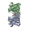 8erpC M: map data used to model this data C: citing same article ( |
|---|---|
| Similar structure data |

-Links
 PDBj
PDBj- Assembly
| Deposited unit | 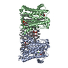
|
|---|---|
| 1 |
|
-Components
| #1: Protein | Mass: 44542.230 Da / Num. of mol.: 2 Source method: isolated from a genetically manipulated source Source: (gene. exp.) Xenopus laevis (African clawed frog) / Gene: chpt1 / Production host:   Spodoptera frugiperda (fall armyworm) Spodoptera frugiperda (fall armyworm)References: UniProt: Q4KLV1, diacylglycerol cholinephosphotransferase#2: Chemical | ChemComp-MG /   Mass: 24.305 Da / Num. of mol.: 4 / Source method: obtained synthetically / Formula: Mg Mass: 24.305 Da / Num. of mol.: 4 / Source method: obtained synthetically / Formula: Mg#3: Chemical | ChemComp-LBN / POPC  Mass: 760.076 Da / Num. of mol.: 26 / Source method: obtained synthetically / Formula: C42H82NO8P / Comment: phospholipid*YM Mass: 760.076 Da / Num. of mol.: 26 / Source method: obtained synthetically / Formula: C42H82NO8P / Comment: phospholipid*YM#4: Chemical | Cytidine diphosphate  Mass: 403.176 Da / Num. of mol.: 2 / Source method: obtained synthetically / Formula: C9H15N3O11P2 / Feature type: SUBJECT OF INVESTIGATION Mass: 403.176 Da / Num. of mol.: 2 / Source method: obtained synthetically / Formula: C9H15N3O11P2 / Feature type: SUBJECT OF INVESTIGATIONHas ligand of interest | Y | |
|---|
-Experimental details
-Experiment
| Experiment | Method: ELECTRON MICROSCOPY |
|---|---|
| EM experiment | Aggregation state: PARTICLE / 3D reconstruction method: single particle reconstruction |
- Sample preparation
| Component | Name: homodimer of choline phosphotransferase 1 / Type: COMPLEX / Entity ID: #1 / Source: RECOMBINANT |
|---|---|
| Source (natural) | Organism: Xenopus laevis (African clawed frog) |
| Source (recombinant) | Organism: Spodoptera frugiperda (fall armyworm) |
| Buffer solution | pH: 7.5 |
| Specimen | Embedding applied: NO / Shadowing applied: NO / Staining applied: NO / Vitrification applied: YES |
| Vitrification | Cryogen name: ETHANE |
- Electron microscopy imaging
| Experimental equipment |  Model: Titan Krios / Image courtesy: FEI Company |
|---|---|
| Microscopy | Model: TFS KRIOS |
| Electron gun | Electron source: FIELD EMISSION GUN / Accelerating voltage: 300 kV / Illumination mode: SPOT SCAN |
| Electron lens | Mode: BRIGHT FIELDBright-field microscopy / Nominal defocus max: 20000 nm / Nominal defocus min: 8000 nm |
| Image recording | Electron dose: 50 e/Å2 / Film or detector model: GATAN K3 BIOQUANTUM (6k x 4k) |
- Processing
| Software | Name: PHENIX / Version: 1.19.1_4122: / Classification: refinement | ||||||||||||||||||||||||
|---|---|---|---|---|---|---|---|---|---|---|---|---|---|---|---|---|---|---|---|---|---|---|---|---|---|
| CTF correction | Type: PHASE FLIPPING AND AMPLITUDE CORRECTION | ||||||||||||||||||||||||
| Symmetry | Point symmetry: C2 (2 fold cyclic) | ||||||||||||||||||||||||
| 3D reconstruction | Resolution: 3.2 Å / Resolution method: FSC 0.143 CUT-OFF / Num. of particles: 465752 / Symmetry type: POINT | ||||||||||||||||||||||||
| Refine LS restraints |
|
