[English] 日本語
Yorodumi
- PDB-8erm: Crystal structure of FliC D2/D3 domains from Pseudomonas aerugino... -

+
Open data


ID or keywords:

Loading...

-
Basic information

Entry
Database: PDB / ID: 8erm
TitleCrystal structure of FliC D2/D3 domains from Pseudomonas aeruginosa PAO1
ComponentsB-type flagellin
KeywordsCELL ADHESION / Flagellin / motility / bacteria / flagella
Function / homology
Function and homology information


bacterial-type flagellum / bacterial-type flagellum-dependent cell motility / structural molecule activity / extracellular region
Similarity search - Function
Flagellin hook, IN motif / Flagellin hook IN motif / Flagellin, C-terminal domain, subdomain 2 / Flagellin, C-terminal domain / Bacterial flagellin C-terminal helical region / Flagellin / Flagellin, N-terminal domain / Bacterial flagellin N-terminal helical region
Similarity search - Domain/homology
Biological speciesPseudomonas aeruginosa PAO1 (bacteria)
MethodX-RAY DIFFRACTION / SYNCHROTRON / MOLECULAR REPLACEMENT / Resolution: 1.475 Å
AuthorsNedeljkovic, M. / Bonsor, D.A. / Postel, S. / Sundberg, E.J.
Funding support1items
OrganizationGrant numberCountry
Not funded
Citation
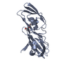
Journal: PLoS Pathog / Year: 2023
Title: An unbroken network of interactions connecting flagellin domains is required for motility in viscous environments.
Authors: Marko Nedeljković / Mark A B Kreutzberger / Sandra Postel / Daniel Bonsor / Yingying Xing / Neil Jacob / William J Schuler / Edward H Egelman / Eric J Sundberg /
Abstract: In its simplest form, bacterial flagellar filaments are composed of flagellin proteins with just two helical inner domains, which together comprise the filament core. Although this minimal filament ...In its simplest form, bacterial flagellar filaments are composed of flagellin proteins with just two helical inner domains, which together comprise the filament core. Although this minimal filament is sufficient to provide motility in many flagellated bacteria, most bacteria produce flagella composed of flagellin proteins with one or more outer domains arranged in a variety of supramolecular architectures radiating from the inner core. Flagellin outer domains are known to be involved in adhesion, proteolysis and immune evasion but have not been thought to be required for motility. Here we show that in the Pseudomonas aeruginosa PAO1 strain, a bacterium that forms a ridged filament with a dimerization of its flagellin outer domains, motility is categorically dependent on these flagellin outer domains. Moreover, a comprehensive network of intermolecular interactions connecting the inner domains to the outer domains, the outer domains to one another, and the outer domains back to the inner domain filament core, is required for motility. This inter-domain connectivity confers PAO1 flagella with increased stability, essential for its motility in viscous environments. Additionally, we find that such ridged flagellar filaments are not unique to Pseudomonas but are, instead, present throughout diverse bacterial phyla.
History
DepositionOct 12, 2022Deposition site: RCSB / Processing site: RCSB
Revision 1.0May 17, 2023Provider: repository / Type: Initial release
Revision 1.1Jun 14, 2023Group: Database references / Category: citation / citation_author
Item: _citation.journal_volume / _citation.page_first ..._citation.journal_volume / _citation.page_first / _citation.page_last / _citation.pdbx_database_id_DOI / _citation.pdbx_database_id_PubMed / _citation.title / _citation_author.identifier_ORCID / _citation_author.name
Revision 1.2Apr 3, 2024Group: Data collection / Refinement description
Category: chem_comp_atom / chem_comp_bond / pdbx_initial_refinement_model

-
Structure visualization

Structure viewerMolecule:
MolmilJmol/JSmol

Downloads & links

-
Assembly

Deposited unit
A: B-type flagellin
B: B-type flagellin
hetero molecules


Theoretical massNumber of molelcules
Total (without water)42,2146
Polymers41,8382
Non-polymers3764
Water6,233346
1
A: B-type flagellin
hetero molecules


Theoretical massNumber of molelcules
Total (without water)21,1033
Polymers20,9191
Non-polymers1842
Water181
TypeNameSymmetry operationNumber
identity operation1_555x,y,z1
MethodPISA
2
B: B-type flagellin
hetero molecules


Theoretical massNumber of molelcules
Total (without water)21,1113
Polymers20,9191
Non-polymers1922
Water181
TypeNameSymmetry operationNumber
identity operation1_555x,y,z1
MethodPISA
Unit cell
Length a, b, c (Å)53.486, 36.492, 87.452
Angle α, β, γ (deg.)90.000, 97.298, 90.000
Int Tables number4
Space group name H-MP1211

-
Components

#1: Protein B-type flagellin


Mass: 20918.945 Da / Num. of mol.: 2
Source method: isolated from a genetically manipulated source
Source: (gene. exp.) Pseudomonas aeruginosa PAO1 (bacteria)
Strain: ATCC 15692 / DSM 22644 / CIP 104116 / JCM 14847 / LMG 12228 / 1C / PRS 101 / PAO1
Gene: fliC, PA1092 / Production host: Escherichia coli (E. coli) / References: UniProt: P72151
#2: Chemical ChemComp-GOL / GLYCEROL / GLYCERIN / PROPANE-1,2,3-TRIOL / Glycerol


Mass: 92.094 Da / Num. of mol.: 2 / Source method: obtained synthetically / Formula: C3H8O3
#3: Chemical ChemComp-SO4 / SULFATE ION / Sulfate


Mass: 96.063 Da / Num. of mol.: 2 / Source method: obtained synthetically / Formula: SO4
#4: Water ChemComp-HOH / water / Water


Mass: 18.015 Da / Num. of mol.: 346 / Source method: isolated from a natural source / Formula: H2O
Has ligand of interestN

-
Experimental details

-
Experiment

ExperimentMethod: X-RAY DIFFRACTION / Number of used crystals: 1

-
Sample preparation

CrystalDensity Matthews: 2.02 Å3/Da / Density % sol: 39.21 %
Crystal growTemperature: 298 K / Method: vapor diffusion, hanging drop / Details: 2 M ammonium sulfate and 0.1 M Tris pH 7.5

-
Data collection

DiffractionMean temperature: 100 K / Serial crystal experiment: N
Diffraction sourceSource: SYNCHROTRON / Site: SSRL / Beamline: BL12-2 / Wavelength: 0.97946 Å
DetectorType: DECTRIS PILATUS 6M / Detector: PIXEL / Date: May 29, 2018
RadiationProtocol: SINGLE WAVELENGTH / Monochromatic (M) / Laue (L): M / Scattering type: x-ray
Radiation wavelengthWavelength: 0.97946 Å / Relative weight: 1
ReflectionResolution: 1.475→36.49 Å / Num. obs: 55941 / % possible obs: 98.4 % / Redundancy: 6.7 % / CC1/2: 0.998 / Rmerge(I) obs: 0.106 / Rpim(I) all: 0.04 / Net I/σ(I): 10.3
Reflection shellResolution: 1.475→1.5 Å / Rmerge(I) obs: 1.008 / Mean I/σ(I) obs: 1.5 / Num. unique obs: 2567 / CC1/2: 0.724 / Rpim(I) all: 0.461

-
Processing

Software
NameVersionClassification
REFMAC5.8.0258refinement
XDSdata reduction
XDSdata scaling
MOLREPphasing
RefinementMethod to determine structure: MOLECULAR REPLACEMENT
Starting model: low resolution SeMet protein

Resolution: 1.475→31.67 Å / Cor.coef. Fo:Fc: 0.966 / Cor.coef. Fo:Fc free: 0.957 / Cross valid method: FREE R-VALUE / ESU R: 0.074 / ESU R Free: 0.074
Details: Hydrogens have been added in their riding positions
RfactorNum. reflection% reflection
Rfree0.202 2773 5 %
Rwork0.1758 53154 -
all0.178 --
obs0.17707 53148 98.22 %
Solvent computationIon probe radii: 0.8 Å / Shrinkage radii: 0.8 Å / VDW probe radii: 1.2 Å / Solvent model: MASK BULK SOLVENT
Displacement parametersBiso mean: 17.579 Å2
Baniso -1Baniso -2Baniso -3
1--0.985 Å2-0 Å20.367 Å2
2--1.758 Å2-0 Å2
3----0.839 Å2
Refinement stepCycle: LAST / Resolution: 1.475→31.67 Å
ProteinNucleic acidLigandSolventTotal
Num. atoms2714 0 22 346 3082
Refine LS restraints
Refine-IDTypeDev idealDev ideal targetNumber
X-RAY DIFFRACTIONr_bond_refined_d0.0170.0132778
X-RAY DIFFRACTIONr_bond_other_d0.0390.0172581
X-RAY DIFFRACTIONr_angle_refined_deg1.891.6283788
X-RAY DIFFRACTIONr_angle_other_deg2.5361.5725988
X-RAY DIFFRACTIONr_dihedral_angle_1_deg6.0515410
X-RAY DIFFRACTIONr_dihedral_angle_2_deg40.06126.22290
X-RAY DIFFRACTIONr_dihedral_angle_3_deg10.42215410
X-RAY DIFFRACTIONr_dihedral_angle_4_deg20.705155
X-RAY DIFFRACTIONr_chiral_restr0.1010.2423
X-RAY DIFFRACTIONr_gen_planes_refined0.0050.023235
X-RAY DIFFRACTIONr_gen_planes_other0.0060.02492
X-RAY DIFFRACTIONr_nbd_refined0.2030.2418
X-RAY DIFFRACTIONr_symmetry_nbd_other0.2130.22269
X-RAY DIFFRACTIONr_nbtor_refined0.1640.21418
X-RAY DIFFRACTIONr_symmetry_nbtor_other0.0840.21466
X-RAY DIFFRACTIONr_xyhbond_nbd_refined0.2570.2231
X-RAY DIFFRACTIONr_symmetry_nbd_refined0.1860.222
X-RAY DIFFRACTIONr_nbd_other0.2850.265
X-RAY DIFFRACTIONr_symmetry_xyhbond_nbd_refined0.2420.235
X-RAY DIFFRACTIONr_mcbond_it1.6311.6081616
X-RAY DIFFRACTIONr_mcbond_other1.6221.6071615
X-RAY DIFFRACTIONr_mcangle_it2.1792.412019
X-RAY DIFFRACTIONr_mcangle_other2.1792.4122020
X-RAY DIFFRACTIONr_scbond_it3.0221.8811162
X-RAY DIFFRACTIONr_scbond_other2.9491.8591155
X-RAY DIFFRACTIONr_scangle_it4.3242.7091764
X-RAY DIFFRACTIONr_scangle_other4.2042.6761753
X-RAY DIFFRACTIONr_lrange_it5.04421.0962907
X-RAY DIFFRACTIONr_lrange_other5.04621.1032908
LS refinement shell
Resolution (Å)Rfactor RfreeNum. reflection RfreeRfactor RworkNum. reflection RworkRefine-ID% reflection obs (%)
1.475-1.5130.3081880.2823684X-RAY DIFFRACTION92.6759
1.513-1.5550.2741830.2423798X-RAY DIFFRACTION98.2963
1.555-1.60.2731700.2233738X-RAY DIFFRACTION98.7867
1.6-1.6490.2452070.2163629X-RAY DIFFRACTION99.0702
1.649-1.7030.2541850.2063487X-RAY DIFFRACTION99.1628
1.703-1.7630.2371890.1923403X-RAY DIFFRACTION99.1444
1.763-1.8290.241950.1813239X-RAY DIFFRACTION98.1423
1.829-1.9040.2131600.173113X-RAY DIFFRACTION96.863
1.904-1.9890.2051610.1663003X-RAY DIFFRACTION99.0607
1.989-2.0860.1991610.1662916X-RAY DIFFRACTION99.3221
2.086-2.1980.2021460.172754X-RAY DIFFRACTION99.1114
2.198-2.3320.1961240.1592662X-RAY DIFFRACTION99.4645
2.332-2.4920.2021190.1572476X-RAY DIFFRACTION99.1973
2.492-2.6920.2021180.1632247X-RAY DIFFRACTION96.7676
2.692-2.9490.1981170.1722131X-RAY DIFFRACTION99.2494
2.949-3.2960.191950.1731928X-RAY DIFFRACTION99.2153
3.296-3.8050.164870.1641722X-RAY DIFFRACTION98.4222
3.805-4.6580.157810.1481428X-RAY DIFFRACTION98.563
4.658-6.5780.192560.191122X-RAY DIFFRACTION97.3554
6.578-31.670.164320.195674X-RAY DIFFRACTION99.1573

+
About Yorodumi

-
News

-
Feb 9, 2022. New format data for meta-information of EMDB entries

New format data for meta-information of EMDB entries

  • Version 3 of the EMDB header file is now the official format.
  • The previous official version 1.9 will be removed from the archive.

Related info.:EMDB header

External links:wwPDB to switch to version 3 of the EMDB data model

-
Aug 12, 2020. Covid-19 info

Covid-19 info

URL: https://pdbj.org/emnavi/covid19.php

New page: Covid-19 featured information page in EM Navigator.

Related info.:Covid-19 info / Mar 5, 2020. Novel coronavirus structure data

+
Mar 5, 2020. Novel coronavirus structure data

Novel coronavirus structure data

Related info.:Yorodumi Speices / Aug 12, 2020. Covid-19 info

External links:COVID-19 featured content - PDBj / Molecule of the Month (242):Coronavirus Proteases

+
Jan 31, 2019. EMDB accession codes are about to change! (news from PDBe EMDB page)

EMDB accession codes are about to change! (news from PDBe EMDB page)

  • The allocation of 4 digits for EMDB accession codes will soon come to an end. Whilst these codes will remain in use, new EMDB accession codes will include an additional digit and will expand incrementally as the available range of codes is exhausted. The current 4-digit format prefixed with “EMD-” (i.e. EMD-XXXX) will advance to a 5-digit format (i.e. EMD-XXXXX), and so on. It is currently estimated that the 4-digit codes will be depleted around Spring 2019, at which point the 5-digit format will come into force.
  • The EM Navigator/Yorodumi systems omit the EMD- prefix.

Related info.:Q: What is EMD? / ID/Accession-code notation in Yorodumi/EM Navigator

External links:EMDB Accession Codes are Changing Soon! / Contact to PDBj

+
Jul 12, 2017. Major update of PDB

Major update of PDB

  • wwPDB released updated PDB data conforming to the new PDBx/mmCIF dictionary.
  • This is a major update changing the version number from 4 to 5, and with Remediation, in which all the entries are updated.
  • In this update, many items about electron microscopy experimental information are reorganized (e.g. em_software).
  • Now, EM Navigator and Yorodumi are based on the updated data.

External links:wwPDB Remediation / Enriched Model Files Conforming to OneDep Data Standards Now Available in the PDB FTP Archive

-
Yorodumi

Thousand views of thousand structures

  • Yorodumi is a browser for structure data from EMDB, PDB, SASBDB, etc.
  • This page is also the successor to EM Navigator detail page, and also detail information page/front-end page for Omokage search.
  • The word "yorodu" (or yorozu) is an old Japanese word meaning "ten thousand". "mi" (miru) is to see.

Related info.:EMDB / PDB / SASBDB / Comparison of 3 databanks / Yorodumi Search / Aug 31, 2016. New EM Navigator & Yorodumi / Yorodumi Papers / Jmol/JSmol / Function and homology information / Changes in new EM Navigator and Yorodumi

Read more