 Movie
Movie Controller


+ Open data

- Basic information
| Entry | Database: PDB / ID: 8emz | ||||||
|---|---|---|---|---|---|---|---|
| Title | Structure of GII.17 norovirus in complex with Nanobody 2 | ||||||
 Components Components |
| ||||||
 Keywords Keywords |  VIRAL PROTEIN / norovirus / Nanobody / inhibitor VIRAL PROTEIN / norovirus / Nanobody / inhibitor | ||||||
| Function / homology | Calicivirus coat protein C-terminal / Calicivirus coat protein C-terminal / Calicivirus coat protein / Calicivirus coat protein / Picornavirus/Calicivirus coat protein / Viral coat protein subunit / VP1 Function and homology information Function and homology information | ||||||
| Biological species |  Norovirus Norovirus Vicugna pacos (alpaca) Vicugna pacos (alpaca) | ||||||
| Method | X-RAY DIFFRACTION / SYNCHROTRON / MOLECULAR REPLACEMENT / Resolution: 1.4 Å | ||||||
 Authors Authors | Kher, G. / Sabin, C. / Pancera, M. / Koromyslova, A. / Hansman, G. | ||||||
| Funding support |  Germany, 1items Germany, 1items
| ||||||
 Citation Citation | Journal: J.Virol. / Year: 2023 Title: Direct Blockade of the Norovirus Histo-Blood Group Antigen Binding Pocket by Nanobodies. Authors: Kher, G. / Sabin, C. / Lun, J.H. / Devant, J.M. / Ruoff, K. / Koromyslova, A.D. / von Itzstein, M. / Pancera, M. / Hansman, G.S. | ||||||
| History |
|
- Structure visualization
| Structure viewer | Molecule: MolmilJmol/JSmol |
|---|
- Downloads & links
-Download
| PDBx/mmCIF format | 8emz.cif.gz | 109.5 KB | Display | PDBx/mmCIF format |
|---|---|---|---|---|
| PDB format | pdb8emz.ent.gz | 81.2 KB | Display | PDB format |
| PDBx/mmJSON format | 8emz.json.gz | Tree view | PDBx/mmJSON format | |
| Others |  Other downloads Other downloads |
-Validation report
| Arichive directory | https://data.pdbj.org/pub/pdb/validation_reports/em/8emzftp://data.pdbj.org/pub/pdb/validation_reports/em/8emz | HTTPS FTP |
|---|
-Related structure data
| Related structure data | 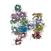 8emyC 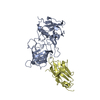 8en0C 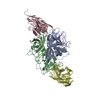 8en1C 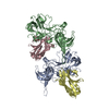 8en2C 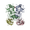 8en3C  8en4C 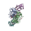 8en5C  8en6C 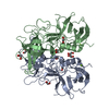 5f4oS S: Starting model for refinement C: citing same article ( |
|---|---|
| Similar structure data |
-Links
 PDBj
PDBj


- Assembly
| Deposited unit | 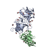
| ||||||||
|---|---|---|---|---|---|---|---|---|---|
| 1 |
| ||||||||
| Unit cell |
| ||||||||
| Components on special symmetry positions |
|
-Components
| #1: Protein | Mass: 34041.898 Da / Num. of mol.: 1 Source method: isolated from a genetically manipulated source Source: (gene. exp.) Norovirus / Production host:  Escherichia coli (E. coli) / References: UniProt: A0A0S1Z370 Escherichia coli (E. coli) / References: UniProt: A0A0S1Z370 | ||||
|---|---|---|---|---|---|
| #2: Antibody | Single-domain antibody Mass: 14128.556 Da / Num. of mol.: 1 Source method: isolated from a genetically manipulated source Source: (gene. exp.) Vicugna pacos (alpaca) / Production host: Escherichia coli (E. coli) | ||||
| #3: Chemical | ChemComp-EDO / Ethylene glycol  Mass: 62.068 Da / Num. of mol.: 16 / Source method: obtained synthetically / Formula: C2H6O2 Mass: 62.068 Da / Num. of mol.: 16 / Source method: obtained synthetically / Formula: C2H6O2#4: Water | ChemComp-HOH / | Water Mass: 18.015 Da / Num. of mol.: 286 / Source method: isolated from a natural source / Formula: H2O Mass: 18.015 Da / Num. of mol.: 286 / Source method: isolated from a natural source / Formula: H2OHas ligand of interest | N | |
-Experimental details
-Experiment
| Experiment | Method: X-RAY DIFFRACTION / Number of used crystals: 1 |
|---|
- Sample preparation
| Crystal | Density Matthews: 2.33 Å3/Da / Density % sol: 47.13 % |
|---|---|
| Crystal grow | Temperature: 291.15 K / Method: vapor diffusion, hanging drop / Details: 0.1 M bicine [pH 9] and 30% [w/v] PEG6000 |
-Data collection
| Diffraction | Mean temperature: 100 K / Serial crystal experiment: N |
|---|---|
| Diffraction source | Source: SYNCHROTRON / Site: SSRL  / Beamline: BL14-1 / Wavelength: 0.9762 Å / Beamline: BL14-1 / Wavelength: 0.9762 Å |
| Detector | Type: DECTRIS EIGER X 16M / Detector: PIXEL / Date: Oct 19, 2020 |
| Radiation | Protocol: SINGLE WAVELENGTH / Monochromatic (M) / Laue (L): M / Scattering type: x-ray |
| Radiation wavelength | Wavelength: 0.9762 Å / Relative weight: 1 |
| Reflection | Resolution: 1.4→117.53 Å / Num. obs: 85355 / % possible obs: 98.5 % / Redundancy: 7.009 % / Biso Wilson estimate: 20.02 Å2 / CC1/2: 1 / Net I/σ(I): 19.1 |
| Reflection shell | Resolution: 2.97→4.19 Å / Redundancy: 7.015 % / Num. unique obs: 2485 / CC1/2: 1 |
- Processing
| Software |
| |||||||||||||||||||||||||||||||||||||||||||||||||||||||||||||||||||||||||||||||||||||||||||||||||||||||||
|---|---|---|---|---|---|---|---|---|---|---|---|---|---|---|---|---|---|---|---|---|---|---|---|---|---|---|---|---|---|---|---|---|---|---|---|---|---|---|---|---|---|---|---|---|---|---|---|---|---|---|---|---|---|---|---|---|---|---|---|---|---|---|---|---|---|---|---|---|---|---|---|---|---|---|---|---|---|---|---|---|---|---|---|---|---|---|---|---|---|---|---|---|---|---|---|---|---|---|---|---|---|---|---|---|---|---|
| Refinement | Method to determine structure: MOLECULAR REPLACEMENT Starting model: 5f4o Resolution: 1.4→48.85 Å / SU ML: 0.19 / Cross valid method: THROUGHOUT / σ(F): 1.35 / Phase error: 26.29 / Stereochemistry target values: ML
| |||||||||||||||||||||||||||||||||||||||||||||||||||||||||||||||||||||||||||||||||||||||||||||||||||||||||
| Solvent computation | Shrinkage radii: 0.9 Å / VDW probe radii: 1.11 Å / Solvent model: FLAT BULK SOLVENT MODEL | |||||||||||||||||||||||||||||||||||||||||||||||||||||||||||||||||||||||||||||||||||||||||||||||||||||||||
| Displacement parameters | Biso max: 97.42 Å2 / Biso mean: 28.5252 Å2 / Biso min: 12.75 Å2 | |||||||||||||||||||||||||||||||||||||||||||||||||||||||||||||||||||||||||||||||||||||||||||||||||||||||||
| Refinement step | Cycle: final / Resolution: 1.4→48.85 Å
| |||||||||||||||||||||||||||||||||||||||||||||||||||||||||||||||||||||||||||||||||||||||||||||||||||||||||
| LS refinement shell | Refine-ID: X-RAY DIFFRACTION / Rfactor Rfree error: 0 / Total num. of bins used: 14
|
