 Movie
Movie Controller

[English] 日本語
 Yorodumi
- PDB-8e0v: DAHP (3-deoxy-D-arabinoheptulosonate-7-phosphate) Synthase comple... -
+ Open data

- Basic information
| Entry | Database: PDB / ID: 8e0v | ||||||||||||||||||
|---|---|---|---|---|---|---|---|---|---|---|---|---|---|---|---|---|---|---|---|
| Title | DAHP (3-deoxy-D-arabinoheptulosonate-7-phosphate) Synthase complexed with Mn(II), PEP, and Pi in unbound:(bound)2:other Conformations | ||||||||||||||||||
 Components Components | Phospho-2-dehydro-3-deoxyheptonate aldolase, Phe-sensitive | ||||||||||||||||||
 Keywords Keywords |  LYASE / DAHP synthase inhibitor / DAHP oxime complex LYASE / DAHP synthase inhibitor / DAHP oxime complex | ||||||||||||||||||
| Function / homology |  Function and homology information3-deoxy-7-phosphoheptulonate synthase / 3-deoxy-7-phosphoheptulonate synthase activity / chorismate biosynthetic process / aromatic amino acid family biosynthetic process / amino acid biosynthetic process / identical protein binding / cytosol / cytoplasm Function and homology information3-deoxy-7-phosphoheptulonate synthase / 3-deoxy-7-phosphoheptulonate synthase activity / chorismate biosynthetic process / aromatic amino acid family biosynthetic process / amino acid biosynthetic process / identical protein binding / cytosol / cytoplasmSimilarity search - Function | ||||||||||||||||||
| Biological species |  Escherichia coli K-12 (bacteria) Escherichia coli K-12 (bacteria) | ||||||||||||||||||
| Method | X-RAY DIFFRACTION / SYNCHROTRON / MOLECULAR REPLACEMENT / Resolution: 2.3 Å | ||||||||||||||||||
 Authors Authors | Berti, P.J. / Junop, M.S. / Grainger, R. | ||||||||||||||||||
| Funding support |  Canada, 5items Canada, 5items
| ||||||||||||||||||
 Citation Citation | Journal: Biochemistry / Year: 2022 Title: Role of Half-of-Sites Reactivity and Inter-Subunit Communications in DAHP Synthase Catalysis and Regulation. Authors: Balachandran, N. / Grainger, R.A. / Rob, T. / Liuni, P. / Wilson, D.J. / Junop, M.S. / Berti, P.J. | ||||||||||||||||||
| History |
|
- Structure visualization
| Structure viewer | Molecule: MolmilJmol/JSmol |
|---|
- Downloads & links
-Download
| PDBx/mmCIF format | 8e0v.cif.gz | 571.3 KB | Display | PDBx/mmCIF format |
|---|---|---|---|---|
| PDB format | pdb8e0v.ent.gz | 448.2 KB | Display | PDB format |
| PDBx/mmJSON format | 8e0v.json.gz | Tree view | PDBx/mmJSON format | |
| Others |  Other downloads Other downloads |
-Validation report
| Arichive directory | https://data.pdbj.org/pub/pdb/validation_reports/e0/8e0vftp://data.pdbj.org/pub/pdb/validation_reports/e0/8e0v | HTTPS FTP |
|---|
-Related structure data
| Related structure data | 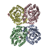 8e0sC 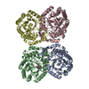 8e0tC 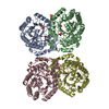 8e0uC 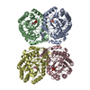 8e0xC 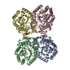 8e0yC 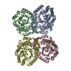 8e0zC 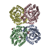 5cksS S: Starting model for refinement C: citing same article ( |
|---|---|
| Similar structure data |
-Links
 PDBj
PDBj


- Assembly
| Deposited unit | 
| ||||||||||||
|---|---|---|---|---|---|---|---|---|---|---|---|---|---|
| 1 |
| ||||||||||||
| Unit cell |
|
-Components
-Protein / Sugars , 2 types, 7 molecules ABCD
| #1: Protein | Mass: 38116.555 Da / Num. of mol.: 4 Source method: isolated from a genetically manipulated source Source: (gene. exp.) Escherichia coli K-12 (bacteria) / Strain: K12 / Gene: aroG, b0754, JW0737 / Production host: Escherichia coli BL21(DE3) (bacteria) / Strain (production host): BL21(DE3)References: UniProt: P0AB91, 3-deoxy-7-phosphoheptulonate synthase#3: Sugar | Galactose Type: D-saccharide, beta linking / Mass: 180.156 Da / Num. of mol.: 3 / Source method: obtained synthetically / Formula: C6H12O6 Type: D-saccharide, beta linking / Mass: 180.156 Da / Num. of mol.: 3 / Source method: obtained synthetically / Formula: C6H12O6 |
|---|
-Non-polymers , 4 types, 933 molecules 
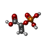


| #2: Chemical | ChemComp-MN /  Mass: 54.938 Da / Num. of mol.: 4 / Source method: obtained synthetically / Formula: Mn / Feature type: SUBJECT OF INVESTIGATION Mass: 54.938 Da / Num. of mol.: 4 / Source method: obtained synthetically / Formula: Mn / Feature type: SUBJECT OF INVESTIGATION#4: Chemical | Phosphoenolpyruvic acid Mass: 168.042 Da / Num. of mol.: 2 / Source method: obtained synthetically / Formula: C3H5O6P / Feature type: SUBJECT OF INVESTIGATION Mass: 168.042 Da / Num. of mol.: 2 / Source method: obtained synthetically / Formula: C3H5O6P / Feature type: SUBJECT OF INVESTIGATION#5: Chemical | Phosphate Mass: 94.971 Da / Num. of mol.: 2 / Source method: obtained synthetically / Formula: PO4 / Feature type: SUBJECT OF INVESTIGATION Mass: 94.971 Da / Num. of mol.: 2 / Source method: obtained synthetically / Formula: PO4 / Feature type: SUBJECT OF INVESTIGATION#6: Water | ChemComp-HOH / | WaterMass: 18.015 Da / Num. of mol.: 925 / Source method: isolated from a natural source / Formula: H2O |
|---|
-Details
| Has ligand of interest | Y |
|---|
-Experimental details
-Experiment
| Experiment | Method: X-RAY DIFFRACTION / Number of used crystals: 1 |
|---|
- Sample preparation
| Crystal | Density Matthews: 2.47 Å3/Da / Density % sol: 50.25 % |
|---|---|
| Crystal grow | Temperature: 277 K / Method: vapor diffusion, hanging drop / pH: 6 Details: 0.1 M trilithium citrate tetrahydrate, 10% (w/v) PEG 3350, 5 mM erythrose 4-phosphate, 5 mM phosphoenolpyruvate, 30% (w/v) D-(+)-galactose |
-Data collection
| Diffraction | Mean temperature: 100 K / Serial crystal experiment: N |
|---|---|
| Diffraction source | Source: SYNCHROTRON / Site: NSLS  / Beamline: X25 / Wavelength: 1.1 Å / Beamline: X25 / Wavelength: 1.1 Å |
| Detector | Type: DECTRIS PILATUS 6M / Detector: PIXEL / Date: Feb 24, 2011 |
| Radiation | Protocol: SINGLE WAVELENGTH / Monochromatic (M) / Laue (L): M / Scattering type: x-ray |
| Radiation wavelength | Wavelength: 1.1 Å / Relative weight: 1 |
| Reflection | Resolution: 2.3→64.45 Å / Num. obs: 66903 / % possible obs: 96.1 % / Observed criterion σ(F): 1.35 / Redundancy: 3.7 % / Biso Wilson estimate: 23.46 Å2 / CC1/2: 0.997 / Rmerge(I) obs: 0.078 / Rpim(I) all: 0.047 / Rrim(I) all: 0.091 / Net I/σ(I): 12.8 |
| Reflection shell | Resolution: 2.3→2.36 Å / Redundancy: 3.6 % / Rmerge(I) obs: 0.391 / Mean I/σ(I) obs: 3.2 / Num. unique obs: 4452 / CC1/2: 0.883 / Rpim(I) all: 0.241 / Rrim(I) all: 0.461 / % possible all: 99.9 |
- Processing
| Software |
| |||||||||||||||||||||||||||||||||||||||||||||||||||||||||||||||||||||||||||||||||||||||||||||||||||||||||
|---|---|---|---|---|---|---|---|---|---|---|---|---|---|---|---|---|---|---|---|---|---|---|---|---|---|---|---|---|---|---|---|---|---|---|---|---|---|---|---|---|---|---|---|---|---|---|---|---|---|---|---|---|---|---|---|---|---|---|---|---|---|---|---|---|---|---|---|---|---|---|---|---|---|---|---|---|---|---|---|---|---|---|---|---|---|---|---|---|---|---|---|---|---|---|---|---|---|---|---|---|---|---|---|---|---|---|
| Refinement | Method to determine structure: MOLECULAR REPLACEMENT Starting model: 5CKS Resolution: 2.3→49.65 Å / SU ML: 0.2247 / Cross valid method: FREE R-VALUE / σ(F): 1.35 / Phase error: 22.457 Stereochemistry target values: GeoStd + Monomer Library + CDL v1.2
| |||||||||||||||||||||||||||||||||||||||||||||||||||||||||||||||||||||||||||||||||||||||||||||||||||||||||
| Solvent computation | Shrinkage radii: 0.9 Å / VDW probe radii: 1.11 Å / Solvent model: FLAT BULK SOLVENT MODEL | |||||||||||||||||||||||||||||||||||||||||||||||||||||||||||||||||||||||||||||||||||||||||||||||||||||||||
| Displacement parameters | Biso mean: 28.15 Å2 | |||||||||||||||||||||||||||||||||||||||||||||||||||||||||||||||||||||||||||||||||||||||||||||||||||||||||
| Refinement step | Cycle: LAST / Resolution: 2.3→49.65 Å
| |||||||||||||||||||||||||||||||||||||||||||||||||||||||||||||||||||||||||||||||||||||||||||||||||||||||||
| Refine LS restraints |
| |||||||||||||||||||||||||||||||||||||||||||||||||||||||||||||||||||||||||||||||||||||||||||||||||||||||||
| LS refinement shell |
|
