Movie
Movie Controller


+ Open data

- Basic information
| Entry | Database: PDB / ID: 8dwv | ||||||
|---|---|---|---|---|---|---|---|
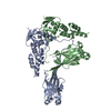
| Title | Full-length wild type SPOP | ||||||
 Components Components | Speckle-type POZ protein | ||||||
 Keywords Keywords |  ONCOPROTEIN / SPOP / ubiquitination / cullin ONCOPROTEIN / SPOP / ubiquitination / cullin | ||||||
| Function / homology |  Function and homology informationregulation of proteolysis / Cul3-RING ubiquitin ligase complex / molecular function inhibitor activity / localization / Hedgehog 'on' state / protein polyubiquitination / proteasome-mediated ubiquitin-dependent protein catabolic process / nuclear speck / ubiquitin protein ligase binding / nucleoplasm ...regulation of proteolysis / Cul3-RING ubiquitin ligase complex / molecular function inhibitor activity / localization / Hedgehog 'on' state / protein polyubiquitination / proteasome-mediated ubiquitin-dependent protein catabolic process / nuclear speck / ubiquitin protein ligase binding / nucleoplasm / nucleus / cytoplasm Function and homology informationregulation of proteolysis / Cul3-RING ubiquitin ligase complex / molecular function inhibitor activity / localization / Hedgehog 'on' state / protein polyubiquitination / proteasome-mediated ubiquitin-dependent protein catabolic process / nuclear speck / ubiquitin protein ligase binding / nucleoplasm ...regulation of proteolysis / Cul3-RING ubiquitin ligase complex / molecular function inhibitor activity / localization / Hedgehog 'on' state / protein polyubiquitination / proteasome-mediated ubiquitin-dependent protein catabolic process / nuclear speck / ubiquitin protein ligase binding / nucleoplasm / nucleus / cytoplasmSimilarity search - Function | ||||||
| Biological species |  Homo sapiens (human) Homo sapiens (human) | ||||||
| Method | ELECTRON MICROSCOPY / single particle reconstruction / cryo EM / Resolution: 3.6 Å | ||||||
 Authors Authors | Cuneo, M.J. / Mittag, T. / O'Flynn, B. / Lo, Y.H. | ||||||
| Funding support |  United States, 1items United States, 1items
| ||||||
 Citation Citation | Journal: Mol Cell / Year: 2023 Title: Higher-order SPOP assembly reveals a basis for cancer mutant dysregulation. Authors: Matthew J Cuneo / Brian G O'Flynn / Yu-Hua Lo / Nafiseh Sabri / Tanja Mittag / Abstract: The speckle-type POZ protein (SPOP) functions in the Cullin3-RING ubiquitin ligase (CRL3) as a receptor for the recognition of substrates involved in cell growth, survival, and signaling. SPOP ...The speckle-type POZ protein (SPOP) functions in the Cullin3-RING ubiquitin ligase (CRL3) as a receptor for the recognition of substrates involved in cell growth, survival, and signaling. SPOP mutations have been attributed to the development of many types of cancers, including prostate and endometrial cancers. Prostate cancer mutations localize in the substrate-binding site of the substrate recognition (MATH) domain and reduce or prevent binding. However, most endometrial cancer mutations are dispersed in seemingly inconspicuous solvent-exposed regions of SPOP, offering no clear basis for their cancer-causing and peculiar gain-of-function properties. Herein, we present the first structure of SPOP in its oligomeric form, uncovering several new interfaces important for SPOP self-assembly and normal function. Given that many previously unaccounted-for cancer mutations are localized in these newly identified interfaces, we uncover molecular mechanisms underlying dysregulation of SPOP function, with effects ranging from gross structural changes to enhanced self-association, and heightened stability and activity. | ||||||
| History |
|
- Structure visualization
| Structure viewer | Molecule: MolmilJmol/JSmol |
|---|
- Downloads & links
-Download
| PDBx/mmCIF format | 8dwv.cif.gz | 352.4 KB | Display | PDBx/mmCIF format |
|---|---|---|---|---|
| PDB format | pdb8dwv.ent.gz | 297.3 KB | Display | PDB format |
| PDBx/mmJSON format | 8dwv.json.gz | Tree view | PDBx/mmJSON format | |
| Others |  Other downloads Other downloads |
-Validation report
| Arichive directory | https://data.pdbj.org/pub/pdb/validation_reports/dw/8dwvftp://data.pdbj.org/pub/pdb/validation_reports/dw/8dwv | HTTPS FTP |
|---|
-Related structure data
| Related structure data |  27761MC  8dwsC 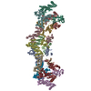 8dwtC  8dwuC M: map data used to model this data C: citing same article ( |
|---|---|
| Similar structure data |


-Links
 PDBj
PDBj


- Assembly
| Deposited unit | 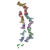
|
|---|---|
| 1 |
|
-Components
| #1: Protein | Mass: 42096.277 Da / Num. of mol.: 6 Source method: isolated from a genetically manipulated source Source: (gene. exp.) Homo sapiens (human) / Gene: SPOP / Production host:  Escherichia coli BL21 (bacteria) / References: UniProt: O43791 Escherichia coli BL21 (bacteria) / References: UniProt: O43791 |
|---|
-Experimental details
-Experiment
| Experiment | Method: ELECTRON MICROSCOPY |
|---|---|
| EM experiment | Aggregation state: FILAMENT / 3D reconstruction method: single particle reconstruction |
- Sample preparation
| Component | Name: SPOP E47K Mutant / Type: COMPLEX / Entity ID: all / Source: RECOMBINANT |
|---|---|
| Molecular weight | Experimental value: NO |
| Source (natural) | Organism: Homo sapiens (human) |
| Source (recombinant) | Organism: Escherichia coli BL21 (bacteria) |
| Buffer solution | pH: 7.5 / Details: 20 mM HEPES pH 7.5, 400 mM NaCl, 5 mM DTT |
| Specimen | Conc.: 1 mg/ml / Embedding applied: NO / Shadowing applied: NO / Staining applied: NO / Vitrification applied: YES |
| Vitrification | Instrument: FEI VITROBOT MARK III / Cryogen name: ETHANE / Humidity: 100 % |
- Electron microscopy imaging
| Experimental equipment |  Model: Titan Krios / Image courtesy: FEI Company |
|---|---|
| Microscopy | Model: FEI TITAN KRIOS |
| Electron gun | Electron source: FIELD EMISSION GUN / Accelerating voltage: 300 kV / Illumination mode: OTHER |
| Electron lens | Mode: BRIGHT FIELDBright-field microscopy / Nominal defocus max: 1800 nm / Nominal defocus min: 600 nm / Alignment procedure: COMA FREE |
| Specimen holder | Cryogen: NITROGEN / Specimen holder model: FEI TITAN KRIOS AUTOGRID HOLDER |
| Image recording | Electron dose: 65 e/Å2 / Film or detector model: GATAN K3 (6k x 4k) |
- Processing
| EM software |
| |||||||||||||||
|---|---|---|---|---|---|---|---|---|---|---|---|---|---|---|---|---|
| CTF correction | Type: PHASE FLIPPING AND AMPLITUDE CORRECTION | |||||||||||||||
| 3D reconstruction | Resolution: 3.6 Å / Resolution method: FSC 0.143 CUT-OFF / Num. of particles: 572000 / Symmetry type: POINT | |||||||||||||||
| Atomic model building | Protocol: OTHER / Space: REAL | |||||||||||||||
| Atomic model building | PDB-ID: 3HQI |