 Movie
Movie Controller

[English] 日本語
 Yorodumi
- PDB-8dir: The complex structure between human IgG1 Fc and its high affinity... -
+ Open data

- Basic information
| Entry | Database: PDB / ID: 8dir | ||||||
|---|---|---|---|---|---|---|---|
| Title | The complex structure between human IgG1 Fc and its high affinity receptor FcgRI H174R variant | ||||||
 Components Components |
| ||||||
 Keywords Keywords |  IMMUNE SYSTEM / High affinity IgG1 Fc receptor IMMUNE SYSTEM / High affinity IgG1 Fc receptor | ||||||
| Function / homology |  Function and homology information Function and homology informationhigh-affinity IgG receptor activity / positive regulation of type III hypersensitivity / phagocytosis, recognition / positive regulation of type IIa hypersensitivity / complement-dependent cytotoxicity / antibody-dependent cellular cytotoxicity / IgG binding / Cross-presentation of soluble exogenous antigens (endosomes) / Fc-gamma receptor I complex binding / Classical antibody-mediated complement activation ...high-affinity IgG receptor activity / positive regulation of type III hypersensitivity / phagocytosis, recognition / positive regulation of type IIa hypersensitivity / complement-dependent cytotoxicity / antibody-dependent cellular cytotoxicity / IgG binding / Cross-presentation of soluble exogenous antigens (endosomes) / Fc-gamma receptor I complex binding / Classical antibody-mediated complement activation / phagocytosis, engulfment / Initial triggering of complement / immunoglobulin complex, circulating / IgG immunoglobulin complex / immunoglobulin receptor binding / antigen processing and presentation of exogenous peptide antigen via MHC class I / FCGR activation / regulation of immune response / Role of phospholipids in phagocytosis / positive regulation of protein tyrosine kinase activity / positive regulation of phagocytosis / complement activation, classical pathway / antigen binding / receptor-mediated endocytosis / FCGR3A-mediated IL10 synthesis / Regulation of Complement cascade / FCGR3A-mediated phagocytosis / B cell receptor signaling pathway / clathrin-coated endocytic vesicle membrane / Regulation of actin dynamics for phagocytic cup formation / Immunoregulatory interactions between a Lymphoid and a non-Lymphoid cell / Interferon gamma signaling / transmembrane signaling receptor activity / antibacterial humoral response / early endosome membrane / Interleukin-4 and Interleukin-13 signaling / blood microparticle / adaptive immune response / cell surface receptor signaling pathway / defense response to bacterium / immune response / external side of plasma membrane / innate immune response / signal transduction / extracellular space / extracellular exosome / extracellular region / plasma membraneSimilarity search - Function | ||||||
| Biological species |  Homo sapiens (human) Homo sapiens (human) | ||||||
| Method | X-RAY DIFFRACTION / SYNCHROTRON / MOLECULAR REPLACEMENT / Resolution: 2.3 Å | ||||||
 Authors Authors | Lu, J. / Sun, P.D. | ||||||
| Funding support |  United States, 1items United States, 1items
| ||||||
 Citation Citation | Journal: Front Immunol / Year: 2023 Title: Fc gamma RI FG-loop functions as a pH sensitive switch for IgG binding and release. Authors: Lu, J. / Spencer, M. / Zou, Z. / Traver, M. / Brzostowski, J. / Sun, P.D. | ||||||
| History |
|
- Structure visualization
| Structure viewer | Molecule: MolmilJmol/JSmol |
|---|
- Downloads & links
-Download
| PDBx/mmCIF format | 8dir.cif.gz | 312.6 KB | Display | PDBx/mmCIF format |
|---|---|---|---|---|
| PDB format | pdb8dir.ent.gz | 252.2 KB | Display | PDB format |
| PDBx/mmJSON format | 8dir.json.gz | Tree view | PDBx/mmJSON format | |
| Others |  Other downloads Other downloads |
-Validation report
| Arichive directory | https://data.pdbj.org/pub/pdb/validation_reports/di/8dirftp://data.pdbj.org/pub/pdb/validation_reports/di/8dir | HTTPS FTP |
|---|
-Related structure data
| Related structure data | 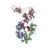 8dinC 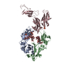 8dj7C  4x4mS S: Starting model for refinement C: citing same article ( |
|---|---|
| Similar structure data |
-Links
 PDBj
PDBj


- Assembly
| Deposited unit | 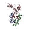
| ||||||||
|---|---|---|---|---|---|---|---|---|---|
| 1 |
| ||||||||
| Unit cell |
|
-Components
| #1: Protein | Mass: 24698.955 Da / Num. of mol.: 2 / Fragment: CH2 and CH3 regions, residues 112-330 Source method: isolated from a genetically manipulated source Source: (gene. exp.) Homo sapiens (human) / Gene: IGHG1 / Production host:   Cricetulus griseus (Chinese hamster) / References: UniProt: P01857 Cricetulus griseus (Chinese hamster) / References: UniProt: P01857#2: Protein | | Mass: 31201.312 Da / Num. of mol.: 1 Source method: isolated from a genetically manipulated source Source: (gene. exp.) Homo sapiens (human) / Gene: FCGR1A, FCG1, FCGR1, IGFR1 / Production host:  Escherichia coli (E. coli) / References: UniProt: P12314 Escherichia coli (E. coli) / References: UniProt: P12314#3: Polysaccharide | / Mass: 1625.490 Da / Num. of mol.: 2Source method: isolated from a genetically manipulated source #4: Chemical |   Mass: 22.990 Da / Num. of mol.: 3 / Source method: obtained synthetically / Formula: Na / Feature type: SUBJECT OF INVESTIGATION Mass: 22.990 Da / Num. of mol.: 3 / Source method: obtained synthetically / Formula: Na / Feature type: SUBJECT OF INVESTIGATION#5: Water | ChemComp-HOH / | Water Mass: 18.015 Da / Num. of mol.: 244 / Source method: isolated from a natural source / Formula: H2O Mass: 18.015 Da / Num. of mol.: 244 / Source method: isolated from a natural source / Formula: H2OHas ligand of interest | Y | |
|---|
-Experimental details
-Experiment
| Experiment | Method: X-RAY DIFFRACTION / Number of used crystals: 1 |
|---|
- Sample preparation
| Crystal | Density Matthews: 3.15 Å3/Da / Density % sol: 60.94 % |
|---|---|
| Crystal grow | Temperature: 293 K / Method: vapor diffusion, hanging drop / Details: 20% PEG3350, 0.2M Sodium Formate (pH6.6) |
-Data collection
| Diffraction | Mean temperature: 100 K / Serial crystal experiment: N |
|---|---|
| Diffraction source | Source: SYNCHROTRON / Site: APS / Beamline: 22-ID / Wavelength: 1 Å |
| Detector | Type: MARMOSAIC 300 mm CCD / Detector: CCD / Date: Jun 3, 2019 |
| Radiation | Protocol: SINGLE WAVELENGTH / Monochromatic (M) / Laue (L): M / Scattering type: x-ray |
| Radiation wavelength | Wavelength: 1 Å / Relative weight: 1 |
| Reflection | Resolution: 2.3→50 Å / Num. obs: 44290 / % possible obs: 99.8 % / Redundancy: 6.5 % / Biso Wilson estimate: 43.45 Å2 / Rmerge(I) obs: 0.07 / Net I/σ(I): 25.3 |
| Reflection shell | Resolution: 2.3→2.34 Å / Rmerge(I) obs: 0.464 / Mean I/σ(I) obs: 2.6 / Num. unique obs: 2235 |
- Processing
| Software |
| |||||||||||||||||||||||||||||||||||||||||||||||||||||||||||||||||||||||||||||||||||||||||||||||||||||||||||||||||||||||
|---|---|---|---|---|---|---|---|---|---|---|---|---|---|---|---|---|---|---|---|---|---|---|---|---|---|---|---|---|---|---|---|---|---|---|---|---|---|---|---|---|---|---|---|---|---|---|---|---|---|---|---|---|---|---|---|---|---|---|---|---|---|---|---|---|---|---|---|---|---|---|---|---|---|---|---|---|---|---|---|---|---|---|---|---|---|---|---|---|---|---|---|---|---|---|---|---|---|---|---|---|---|---|---|---|---|---|---|---|---|---|---|---|---|---|---|---|---|---|---|---|
| Refinement | Method to determine structure: MOLECULAR REPLACEMENT Starting model: 4x4m Resolution: 2.3→36.87 Å / SU ML: 0.31 / Cross valid method: THROUGHOUT / σ(F): 1.36 / Phase error: 24.02 / Stereochemistry target values: ML
| |||||||||||||||||||||||||||||||||||||||||||||||||||||||||||||||||||||||||||||||||||||||||||||||||||||||||||||||||||||||
| Solvent computation | Shrinkage radii: 0.9 Å / VDW probe radii: 1.1 Å / Solvent model: FLAT BULK SOLVENT MODEL | |||||||||||||||||||||||||||||||||||||||||||||||||||||||||||||||||||||||||||||||||||||||||||||||||||||||||||||||||||||||
| Displacement parameters | Biso max: 202.25 Å2 / Biso mean: 61.7028 Å2 / Biso min: 20.93 Å2 | |||||||||||||||||||||||||||||||||||||||||||||||||||||||||||||||||||||||||||||||||||||||||||||||||||||||||||||||||||||||
| Refinement step | Cycle: final / Resolution: 2.3→36.87 Å
| |||||||||||||||||||||||||||||||||||||||||||||||||||||||||||||||||||||||||||||||||||||||||||||||||||||||||||||||||||||||
| LS refinement shell | Refine-ID: X-RAY DIFFRACTION / Rfactor Rfree error: 0 / Total num. of bins used: 16
| |||||||||||||||||||||||||||||||||||||||||||||||||||||||||||||||||||||||||||||||||||||||||||||||||||||||||||||||||||||||
| Refinement TLS params. | Method: refined / Origin x: 34.4459 Å / Origin y: -47.1638 Å / Origin z: 35.6579 Å
| |||||||||||||||||||||||||||||||||||||||||||||||||||||||||||||||||||||||||||||||||||||||||||||||||||||||||||||||||||||||
| Refinement TLS group |
|
