 Movie
Movie Controller


+ Open data

- Basic information
| Entry | Database: PDB / ID: 8dei | ||||||
|---|---|---|---|---|---|---|---|
| Title | Structure of the Cac1 KER domain | ||||||
 Components Components | Maltodextrin-binding protein,Chromatin assembly factor 1 subunit p90 fusion | ||||||
 Keywords Keywords |  DNA BINDING PROTEIN / chromatin / DNA binding DNA BINDING PROTEIN / chromatin / DNA binding | ||||||
| Function / homology |  Function and homology information Function and homology informationCAF-1 complex / DNA replication-dependent chromatin assembly / chromosome, centromeric region / carbohydrate transmembrane transporter activity / nucleosome / nucleosome assembly / chromatin organization / outer membrane-bounded periplasmic space / DNA replication / DNA repair ...CAF-1 complex / DNA replication-dependent chromatin assembly / chromosome, centromeric region / carbohydrate transmembrane transporter activity / nucleosome / nucleosome assembly / chromatin organization / outer membrane-bounded periplasmic space / DNA replication / DNA repair / chromatin / nucleusSimilarity search - Function | ||||||
| Biological species |  Escherichia coli (E. coli) Escherichia coli (E. coli) Saccharomyces cerevisiae (brewer's yeast) Saccharomyces cerevisiae (brewer's yeast) | ||||||
| Method | X-RAY DIFFRACTION / MOLECULAR REPLACEMENT / Resolution: 2.81 Å | ||||||
 Authors Authors | Rosas, R. / Churchill, M.E.A. | ||||||
| Funding support |  United States, 1items United States, 1items
| ||||||
 Citation Citation | Journal: Elife / Year: 2023 Title: A novel single alpha-helix DNA-binding domain in CAF-1 promotes gene silencing and DNA damage survival through tetrasome-length DNA selectivity and spacer function. Authors: Rosas, R. / Aguilar, R.R. / Arslanovic, N. / Seck, A. / Smith, D.J. / Tyler, J.K. / Churchill, M.E.A. | ||||||
| History |
|
- Structure visualization
| Structure viewer | Molecule: MolmilJmol/JSmol |
|---|
- Downloads & links
-Download
| PDBx/mmCIF format | 8dei.cif.gz | 453 KB | Display | PDBx/mmCIF format |
|---|---|---|---|---|
| PDB format | pdb8dei.ent.gz | 298.7 KB | Display | PDB format |
| PDBx/mmJSON format | 8dei.json.gz | Tree view | PDBx/mmJSON format | |
| Others |  Other downloads Other downloads |
-Validation report
| Arichive directory | https://data.pdbj.org/pub/pdb/validation_reports/de/8deiftp://data.pdbj.org/pub/pdb/validation_reports/de/8dei | HTTPS FTP |
|---|
-Related structure data
| Related structure data | 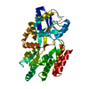 1pebS S: Starting model for refinement |
|---|---|
| Similar structure data |
-Links
 PDBj
PDBj


- Assembly
| Deposited unit | 
| ||||||||||||
|---|---|---|---|---|---|---|---|---|---|---|---|---|---|
| 1 | 
| ||||||||||||
| 2 | 
| ||||||||||||
| 3 | 
| ||||||||||||
| 4 | 
| ||||||||||||
| Unit cell |
|
-Components
| #1: Protein | Mass: 55608.172 Da / Num. of mol.: 4 / Fragment: KER domain, residues 136-225 Source method: isolated from a genetically manipulated source Source: (gene. exp.) Escherichia coli (E. coli), (gene. exp.) Saccharomyces cerevisiae (brewer's yeast)Gene: malE, malE_1, malE_2, RLF2, CAC1, YPR018W, YP9531.12 / Strain: ATCC 204508 / S288c / Production host: Escherichia coli (E. coli) / References: UniProt: C3SHQ8, UniProt: Q12495#2: Chemical | ChemComp-PEG / Diethylene glycol  Mass: 106.120 Da / Num. of mol.: 5 / Source method: obtained synthetically / Formula: C4H10O3 / Feature type: SUBJECT OF INVESTIGATION Mass: 106.120 Da / Num. of mol.: 5 / Source method: obtained synthetically / Formula: C4H10O3 / Feature type: SUBJECT OF INVESTIGATION#3: Chemical | ChemComp-PO4 / | Phosphate  Mass: 94.971 Da / Num. of mol.: 1 / Source method: obtained synthetically / Formula: PO4 Mass: 94.971 Da / Num. of mol.: 1 / Source method: obtained synthetically / Formula: PO4#4: Chemical | ChemComp-GOL / | Glycerol  Mass: 92.094 Da / Num. of mol.: 1 / Source method: obtained synthetically / Formula: C3H8O3 Mass: 92.094 Da / Num. of mol.: 1 / Source method: obtained synthetically / Formula: C3H8O3#5: Water | ChemComp-HOH / | Water Mass: 18.015 Da / Num. of mol.: 11 / Source method: isolated from a natural source / Formula: H2O Mass: 18.015 Da / Num. of mol.: 11 / Source method: isolated from a natural source / Formula: H2OHas ligand of interest | Y | |
|---|
-Experimental details
-Experiment
| Experiment | Method: X-RAY DIFFRACTION / Number of used crystals: 1 |
|---|
- Sample preparation
| Crystal | Density Matthews: 2.3 Å3/Da / Density % sol: 46.46 % |
|---|---|
| Crystal grow | Temperature: 288 K / Method: vapor diffusion, hanging drop / Details: 0.1 M Phosphate/citrate pH 4.2 and 30 % PEG 300 |
-Data collection
| Diffraction | Mean temperature: 100 K / Serial crystal experiment: N |
|---|---|
| Diffraction source | Source: ROTATING ANODE / Type: RIGAKU MICROMAX-007 HF / Wavelength: 1.54 Å |
| Detector | Type: DECTRIS PILATUS 200K / Detector: PIXEL / Date: Mar 30, 2018 |
| Radiation | Protocol: SINGLE WAVELENGTH / Monochromatic (M) / Laue (L): M / Scattering type: x-ray |
| Radiation wavelength | Wavelength: 1.54 Å / Relative weight: 1 |
| Reflection | Resolution: 2.81→28.68 Å / Num. obs: 48320 / % possible obs: 99 % / Redundancy: 3.9 % / Biso Wilson estimate: 46.75 Å2 / CC1/2: 0.979 / Net I/σ(I): 7.3 |
| Reflection shell | Resolution: 2.81→2.91 Å / Redundancy: 3 % / Num. unique obs: 13259 / CC1/2: 0.695 / % possible all: 90.9 |
- Processing
| Software |
| |||||||||||||||||||||||||||||||||||||||||||||||||||||||||||||||||||||||||||||||||||||||||||||||||||||||||||||||||||||||||||||||||||||||||||||||||||||||||||||||||||||||||||||||||||||||||||||||||||||||||||||||||||||||||
|---|---|---|---|---|---|---|---|---|---|---|---|---|---|---|---|---|---|---|---|---|---|---|---|---|---|---|---|---|---|---|---|---|---|---|---|---|---|---|---|---|---|---|---|---|---|---|---|---|---|---|---|---|---|---|---|---|---|---|---|---|---|---|---|---|---|---|---|---|---|---|---|---|---|---|---|---|---|---|---|---|---|---|---|---|---|---|---|---|---|---|---|---|---|---|---|---|---|---|---|---|---|---|---|---|---|---|---|---|---|---|---|---|---|---|---|---|---|---|---|---|---|---|---|---|---|---|---|---|---|---|---|---|---|---|---|---|---|---|---|---|---|---|---|---|---|---|---|---|---|---|---|---|---|---|---|---|---|---|---|---|---|---|---|---|---|---|---|---|---|---|---|---|---|---|---|---|---|---|---|---|---|---|---|---|---|---|---|---|---|---|---|---|---|---|---|---|---|---|---|---|---|---|---|---|---|---|---|---|---|---|---|---|---|---|---|---|---|---|
| Refinement | Method to determine structure: MOLECULAR REPLACEMENT Starting model: 1PEB Resolution: 2.81→28.68 Å / SU ML: 0.4341 / Cross valid method: FREE R-VALUE / σ(F): 1.34 / Phase error: 31.7751 Stereochemistry target values: GeoStd + Monomer Library + CDL v1.2
| |||||||||||||||||||||||||||||||||||||||||||||||||||||||||||||||||||||||||||||||||||||||||||||||||||||||||||||||||||||||||||||||||||||||||||||||||||||||||||||||||||||||||||||||||||||||||||||||||||||||||||||||||||||||||
| Solvent computation | Shrinkage radii: 0.9 Å / VDW probe radii: 1.11 Å / Solvent model: FLAT BULK SOLVENT MODEL | |||||||||||||||||||||||||||||||||||||||||||||||||||||||||||||||||||||||||||||||||||||||||||||||||||||||||||||||||||||||||||||||||||||||||||||||||||||||||||||||||||||||||||||||||||||||||||||||||||||||||||||||||||||||||
| Displacement parameters | Biso mean: 51.56 Å2 | |||||||||||||||||||||||||||||||||||||||||||||||||||||||||||||||||||||||||||||||||||||||||||||||||||||||||||||||||||||||||||||||||||||||||||||||||||||||||||||||||||||||||||||||||||||||||||||||||||||||||||||||||||||||||
| Refinement step | Cycle: LAST / Resolution: 2.81→28.68 Å
| |||||||||||||||||||||||||||||||||||||||||||||||||||||||||||||||||||||||||||||||||||||||||||||||||||||||||||||||||||||||||||||||||||||||||||||||||||||||||||||||||||||||||||||||||||||||||||||||||||||||||||||||||||||||||
| Refine LS restraints |
| |||||||||||||||||||||||||||||||||||||||||||||||||||||||||||||||||||||||||||||||||||||||||||||||||||||||||||||||||||||||||||||||||||||||||||||||||||||||||||||||||||||||||||||||||||||||||||||||||||||||||||||||||||||||||
| LS refinement shell |
|
