 Movie
Movie Controller

[English] 日本語
 Yorodumi
- PDB-8d9p: De Novo Photosynthetic Reaction Center Protein Equipped with Heme... -
+ Open data

- Basic information
| Entry | Database: PDB / ID: 8d9p | ||||||
|---|---|---|---|---|---|---|---|
| Title | De Novo Photosynthetic Reaction Center Protein Equipped with Heme B and Mn(II) cations | ||||||
 Components Components | Reaction center maquette | ||||||
 Keywords Keywords |  DE NOVO PROTEIN / maquette / protein design / charge separation / artificial photosynthesis DE NOVO PROTEIN / maquette / protein design / charge separation / artificial photosynthesis | ||||||
| Function / homology | PROTOPORPHYRIN IX CONTAINING FE / :  Function and homology information Function and homology information | ||||||
| Biological species | synthetic construct (others) | ||||||
| Method | X-RAY DIFFRACTION / MOLECULAR REPLACEMENT / Resolution: 1.9 Å | ||||||
 Authors Authors | Ennist, N.M. / Stayrook, S.E. / Dutton, P.L. / Moser, C.C. | ||||||
| Funding support |  United States, 1items United States, 1items
| ||||||
 Citation Citation | Journal: Front Mol Biosci / Year: 2022 Title: Rational design of photosynthetic reaction center protein maquettes. Authors: Ennist, N.M. / Stayrook, S.E. / Dutton, P.L. / Moser, C.C. #1: Journal: To Be PublishedTitle: De Novo Protein Design of Photochemical Reaction Centers Authors: Ennist, N.M. / Zhao, Z. / Stayrook, S.E. / Discher, B.M. / Dutton, P.L. / Moser, C.C. | ||||||
| History |
|
- Structure visualization
| Structure viewer | Molecule: MolmilJmol/JSmol |
|---|
- Downloads & links
-Download
| PDBx/mmCIF format | 8d9p.cif.gz | 58.7 KB | Display | PDBx/mmCIF format |
|---|---|---|---|---|
| PDB format | pdb8d9p.ent.gz | 40.3 KB | Display | PDB format |
| PDBx/mmJSON format | 8d9p.json.gz | Tree view | PDBx/mmJSON format | |
| Others |  Other downloads Other downloads |
-Validation report
| Arichive directory | https://data.pdbj.org/pub/pdb/validation_reports/d9/8d9pftp://data.pdbj.org/pub/pdb/validation_reports/d9/8d9p | HTTPS FTP |
|---|
-Related structure data
| Related structure data |  8d9oC 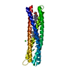 5vjsS S: Starting model for refinement C: citing same article ( |
|---|---|
| Similar structure data |
-Links
 PDBj
PDBj
- Assembly
| Deposited unit | 
| ||||||||||
|---|---|---|---|---|---|---|---|---|---|---|---|
| 1 |
| ||||||||||
| Unit cell |
|
-Components
| #1: Protein | Mass: 22530.430 Da / Num. of mol.: 1 Source method: isolated from a genetically manipulated source Source: (gene. exp.) synthetic construct (others) / Production host:  Escherichia coli (E. coli) Escherichia coli (E. coli) | ||||||
|---|---|---|---|---|---|---|---|
| #2: Chemical | ChemComp-HEM / Heme B  Mass: 616.487 Da / Num. of mol.: 1 / Source method: obtained synthetically / Formula: C34H32FeN4O4 / Feature type: SUBJECT OF INVESTIGATION Mass: 616.487 Da / Num. of mol.: 1 / Source method: obtained synthetically / Formula: C34H32FeN4O4 / Feature type: SUBJECT OF INVESTIGATION | ||||||
| #3: Chemical |   Mass: 54.938 Da / Num. of mol.: 2 / Source method: obtained synthetically / Formula: Mn / Feature type: SUBJECT OF INVESTIGATION Mass: 54.938 Da / Num. of mol.: 2 / Source method: obtained synthetically / Formula: Mn / Feature type: SUBJECT OF INVESTIGATION#4: Chemical | ChemComp-CL / | Chloride  Mass: 35.453 Da / Num. of mol.: 1 / Source method: obtained synthetically / Formula: Cl Mass: 35.453 Da / Num. of mol.: 1 / Source method: obtained synthetically / Formula: Cl#5: Water | ChemComp-HOH / | Water Mass: 18.015 Da / Num. of mol.: 82 / Source method: isolated from a natural source / Formula: H2O Mass: 18.015 Da / Num. of mol.: 82 / Source method: isolated from a natural source / Formula: H2OHas ligand of interest | Y | |
-Experimental details
-Experiment
| Experiment | Method: X-RAY DIFFRACTION / Number of used crystals: 1 |
|---|
- Sample preparation
| Crystal | Density Matthews: 2.68 Å3/Da / Density % sol: 54.11 % / Description: octahedral |
|---|---|
| Crystal grow | Temperature: 277 K / Method: vapor diffusion, hanging drop / pH: 5.9 Details: 1.2 M Li2SO4, 0.5 M (NH4)2SO4, 150 mM Na citrate, pH 5.9 Cryoprotectant: 28% glycerol, 1.25 M Li2SO4, 0.5 M (NH4)2SO4, 100 mM Na citrate, pH 5.85 |
-Data collection
| Diffraction | Mean temperature: 100 K / Serial crystal experiment: N |
|---|---|
| Diffraction source | Source: ROTATING ANODE / Type: RIGAKU MICROMAX-007 HF / Wavelength: 1.54178 Å |
| Detector | Type: RIGAKU SATURN 944+ / Detector: CCD / Date: Sep 23, 2014 / Details: Osmic VariMax mirror |
| Radiation | Protocol: SINGLE WAVELENGTH / Monochromatic (M) / Laue (L): M / Scattering type: x-ray |
| Radiation wavelength | Wavelength: 1.54178 Å / Relative weight: 1 |
| Reflection | Resolution: 1.9→23.25 Å / Num. obs: 20059 / % possible obs: 98.4 % / Redundancy: 12.6 % / Biso Wilson estimate: 28.22 Å2 / CC1/2: 0.999 / Rmerge(I) obs: 0.089 / Rrim(I) all: 0.093 / Net I/σ(I): 22.22 |
| Reflection shell | Resolution: 1.9→1.95 Å / Mean I/σ(I) obs: 1.66 / Num. unique obs: 1271 / CC1/2: 0.619 / % possible all: 86.6 |
- Processing
| Software |
| |||||||||||||||||||||||||||||||||||||||||||||||||||||||||||||||||||||||||||||||||||||||||||||||||||||||||
|---|---|---|---|---|---|---|---|---|---|---|---|---|---|---|---|---|---|---|---|---|---|---|---|---|---|---|---|---|---|---|---|---|---|---|---|---|---|---|---|---|---|---|---|---|---|---|---|---|---|---|---|---|---|---|---|---|---|---|---|---|---|---|---|---|---|---|---|---|---|---|---|---|---|---|---|---|---|---|---|---|---|---|---|---|---|---|---|---|---|---|---|---|---|---|---|---|---|---|---|---|---|---|---|---|---|---|
| Refinement | Method to determine structure: MOLECULAR REPLACEMENT Starting model: 5VJS Resolution: 1.9→23.25 Å / Cor.coef. Fo:Fc: 0.945 / Cor.coef. Fo:Fc free: 0.937 / SU B: 4.475 / SU ML: 0.125 / Cross valid method: FREE R-VALUE / ESU R: 0.16 / ESU R Free: 0.142 / Stereochemistry target values: MAXIMUM LIKELIHOOD Details: HYDROGENS HAVE BEEN ADDED IN THE RIDING POSITIONS U VALUES : REFINED INDIVIDUALLY
| |||||||||||||||||||||||||||||||||||||||||||||||||||||||||||||||||||||||||||||||||||||||||||||||||||||||||
| Solvent computation | Ion probe radii: 1 Å / Shrinkage radii: 1 Å / VDW probe radii: 1.5 Å / Solvent model: MASK | |||||||||||||||||||||||||||||||||||||||||||||||||||||||||||||||||||||||||||||||||||||||||||||||||||||||||
| Displacement parameters | Biso mean: 37.956 Å2
| |||||||||||||||||||||||||||||||||||||||||||||||||||||||||||||||||||||||||||||||||||||||||||||||||||||||||
| Refinement step | Cycle: LAST / Resolution: 1.9→23.25 Å
| |||||||||||||||||||||||||||||||||||||||||||||||||||||||||||||||||||||||||||||||||||||||||||||||||||||||||
| Refine LS restraints |
| |||||||||||||||||||||||||||||||||||||||||||||||||||||||||||||||||||||||||||||||||||||||||||||||||||||||||
| LS refinement shell | Resolution: 1.902→1.951 Å / Total num. of bins used: 20
|
