Movie
Movie Controller


+ Open data

- Basic information
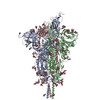
| Entry | Database: PDB / ID: 8d55 | |||||||||||||||||||||
|---|---|---|---|---|---|---|---|---|---|---|---|---|---|---|---|---|---|---|---|---|---|---|
| Title | Closed state of SARS-CoV-2 BA.2 variant spike protein | |||||||||||||||||||||
 Components Components | Spike glycoprotein Spike protein Spike protein | |||||||||||||||||||||
 Keywords Keywords | VIRAL PROTEIN | |||||||||||||||||||||
| Function / homology |  Function and homology information Function and homology informationMaturation of spike protein / viral translation / Translation of Structural Proteins / Virion Assembly and Release / host cell surface / host extracellular space / suppression by virus of host tetherin activity / Induction of Cell-Cell Fusion / structural constituent of virion / host cell endoplasmic reticulum-Golgi intermediate compartment membrane ...Maturation of spike protein / viral translation / Translation of Structural Proteins / Virion Assembly and Release / host cell surface / host extracellular space / suppression by virus of host tetherin activity / Induction of Cell-Cell Fusion / structural constituent of virion / host cell endoplasmic reticulum-Golgi intermediate compartment membrane / entry receptor-mediated virion attachment to host cell / receptor-mediated endocytosis of virus by host cell / Attachment and Entry / membrane fusion / positive regulation of viral entry into host cell / receptor-mediated virion attachment to host cell / receptor ligand activity / host cell surface receptor binding / fusion of virus membrane with host plasma membrane / fusion of virus membrane with host endosome membrane / viral envelope / symbiont-mediated suppression of host type I interferon-mediated signaling pathway / virion attachment to host cell / SARS-CoV-2 activates/modulates innate and adaptive immune responses / host cell plasma membrane / virion membrane / membrane / identical protein binding / plasma membraneSimilarity search - Function | |||||||||||||||||||||
| Biological species |   Severe acute respiratory syndrome coronavirus 2 Severe acute respiratory syndrome coronavirus 2 | |||||||||||||||||||||
| Method | ELECTRON MICROSCOPY / single particle reconstruction / cryo EM / Resolution: 2.8 Å | |||||||||||||||||||||
 Authors Authors | Zhang, J. / Tang, W.C. / Gao, H.L. / Shi, W. / Peng, H.Q. / Volloch, S.R. / Xiao, T.S. / Chen, B. | |||||||||||||||||||||
| Funding support |  United States, 6items United States, 6items
| |||||||||||||||||||||
 Citation Citation | Journal: Nat Struct Mol Biol / Year: 2023 Title: Structural and functional characteristics of the SARS-CoV-2 Omicron subvariant BA.2 spike protein. Authors: Jun Zhang / Weichun Tang / Hailong Gao / Christy L Lavine / Wei Shi / Hanqin Peng / Haisun Zhu / Krishna Anand / Matina Kosikova / Hyung Joon Kwon / Pei Tong / Avneesh Gautam / Sophia Rits- ...Authors: Jun Zhang / Weichun Tang / Hailong Gao / Christy L Lavine / Wei Shi / Hanqin Peng / Haisun Zhu / Krishna Anand / Matina Kosikova / Hyung Joon Kwon / Pei Tong / Avneesh Gautam / Sophia Rits-Volloch / Shaowei Wang / Megan L Mayer / Duane R Wesemann / Michael S Seaman / Jianming Lu / Tianshu Xiao / Hang Xie / Bing Chen / Abstract: The Omicron subvariant BA.2 has become the dominant circulating strain of severe acute respiratory syndrome coronavirus 2 (SARS-CoV-2) in many countries. Here, we have characterized structural, ...The Omicron subvariant BA.2 has become the dominant circulating strain of severe acute respiratory syndrome coronavirus 2 (SARS-CoV-2) in many countries. Here, we have characterized structural, functional and antigenic properties of the full-length BA.2 spike (S) protein and compared replication of the authentic virus in cell culture and an animal model with previously prevalent variants. BA.2 S can fuse membranes slightly more efficiently than Omicron BA.1, but still less efficiently than other previous variants. Both BA.1 and BA.2 viruses replicated substantially faster in animal lungs than the early G614 (B.1) strain in the absence of pre-existing immunity, possibly explaining the increased transmissibility despite their functionally compromised spikes. As in BA.1, mutations in the BA.2 S remodel its antigenic surfaces, leading to strong resistance to neutralizing antibodies. These results suggest that both immune evasion and replicative advantage may contribute to the heightened transmissibility of the Omicron subvariants. | |||||||||||||||||||||
| History |
|
- Structure visualization
| Structure viewer | Molecule: MolmilJmol/JSmol |
|---|
- Downloads & links
-Download
| PDBx/mmCIF format | 8d55.cif.gz | 695.8 KB | Display | PDBx/mmCIF format |
|---|---|---|---|---|
| PDB format | pdb8d55.ent.gz | 573.3 KB | Display | PDB format |
| PDBx/mmJSON format | 8d55.json.gz | Tree view | PDBx/mmJSON format | |
| Others |  Other downloads Other downloads |
-Validation report
| Arichive directory | https://data.pdbj.org/pub/pdb/validation_reports/d5/8d55ftp://data.pdbj.org/pub/pdb/validation_reports/d5/8d55 | HTTPS FTP |
|---|
-Related structure data
| Related structure data |  27205MC 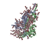 8d56C 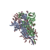 8d5aC M: map data used to model this data C: citing same article ( |
|---|---|
| Similar structure data |


-Links
 PDBj
PDBj


- Assembly
| Deposited unit | 
|
|---|---|
| 1 |
|
-Components
| #1: Protein | Spike protein / S glycoprotein / E2 / Peplomer protein Mass: 144688.156 Da / Num. of mol.: 3 Source method: isolated from a genetically manipulated source Source: (gene. exp.) Severe acute respiratory syndrome coronavirus 2Gene: S, 2 / Variant: BA.2 SARS-CoV-2 spike protein variant / Production host:  Homo sapiens (human) / References: UniProt: P0DTC2 Homo sapiens (human) / References: UniProt: P0DTC2#2: Polysaccharide | alpha-D-mannopyranose-(1-4)-2-acetamido-2-deoxy-beta-D-glucopyranose-(1-4)-2-acetamido-2-deoxy-beta- ...alpha-D-mannopyranose-(1-4)-2-acetamido-2-deoxy-beta-D-glucopyranose-(1-4)-2-acetamido-2-deoxy-beta-D-glucopyranose / Mass: 586.542 Da / Num. of mol.: 21Source method: isolated from a genetically manipulated source #3: Polysaccharide | 2-acetamido-2-deoxy-beta-D-glucopyranose-(1-4)-2-acetamido-2-deoxy-beta-D-glucopyranose / Mass: 424.401 Da / Num. of mol.: 6Source method: isolated from a genetically manipulated source #4: Polysaccharide | / Mass: 570.542 Da / Num. of mol.: 3Source method: isolated from a genetically manipulated source #5: Sugar | ChemComp-NAG / N-Acetylglucosamine  Type: D-saccharide, beta linking / Mass: 221.208 Da / Num. of mol.: 21 / Source method: obtained synthetically / Formula: C8H15NO6 / Feature type: SUBJECT OF INVESTIGATION Type: D-saccharide, beta linking / Mass: 221.208 Da / Num. of mol.: 21 / Source method: obtained synthetically / Formula: C8H15NO6 / Feature type: SUBJECT OF INVESTIGATIONHas ligand of interest | Y | |
|---|
-Experimental details
-Experiment
| Experiment | Method: ELECTRON MICROSCOPY |
|---|---|
| EM experiment | Aggregation state: PARTICLE / 3D reconstruction method: single particle reconstruction |
- Sample preparation
| Component | Name: Closed state of pre-fusion SARS-CoV-2 BA.2 variant spike protein Type: COMPLEX Details: Closed state of pre-fusion SARS-CoV-2 BA.2 variant spike protein Entity ID: #1 / Source: RECOMBINANT | ||||||||||||||||||||
|---|---|---|---|---|---|---|---|---|---|---|---|---|---|---|---|---|---|---|---|---|---|
| Molecular weight | Value: 600 kDa/nm / Experimental value: NO | ||||||||||||||||||||
| Source (natural) | Organism: Severe acute respiratory syndrome coronavirus 2 | ||||||||||||||||||||
| Source (recombinant) | Organism: Homo sapiens (human) | ||||||||||||||||||||
| Buffer solution | pH: 7.5 | ||||||||||||||||||||
| Buffer component |
| ||||||||||||||||||||
| Specimen | Conc.: 1 mg/ml / Embedding applied: NO / Shadowing applied: NO / Staining applied: NO / Vitrification applied: YES | ||||||||||||||||||||
| Vitrification | Instrument: FEI VITROBOT MARK IV / Cryogen name: ETHANE / Humidity: 100 % / Chamber temperature: 277.15 K |
- Electron microscopy imaging
| Experimental equipment |  Model: Titan Krios / Image courtesy: FEI Company |
|---|---|
| Microscopy | Model: FEI TITAN KRIOS |
| Electron gun | Electron source: FIELD EMISSION GUN / Accelerating voltage: 300 kV / Illumination mode: FLOOD BEAM |
| Electron lens | Mode: BRIGHT FIELDBright-field microscopy / Nominal defocus max: 2200 nm / Nominal defocus min: 500 nm |
| Image recording | Electron dose: 53.853 e/Å2 / Film or detector model: GATAN K3 BIOQUANTUM (6k x 4k) |
- Processing
| Software | Name: PHENIX / Version: 1.20_4459: / Classification: refinement | ||||||||||||||||||||||||
|---|---|---|---|---|---|---|---|---|---|---|---|---|---|---|---|---|---|---|---|---|---|---|---|---|---|
| EM software |
| ||||||||||||||||||||||||
| CTF correction | Type: PHASE FLIPPING AND AMPLITUDE CORRECTION | ||||||||||||||||||||||||
| Particle selection | Num. of particles selected: 6763049 | ||||||||||||||||||||||||
| 3D reconstruction | Resolution: 2.8 Å / Resolution method: FSC 0.143 CUT-OFF / Num. of particles: 160774 / Symmetry type: POINT | ||||||||||||||||||||||||
| Atomic model building | Protocol: AB INITIO MODEL | ||||||||||||||||||||||||
| Atomic model building | PDB-ID: 7KRR Pdb chain-ID: A / Pdb chain residue range: 14-1211 | ||||||||||||||||||||||||
| Refine LS restraints |
|