 Movie
Movie Controller


+ Open data

- Basic information
| Entry | Database: PDB / ID: 8czk | ||||||
|---|---|---|---|---|---|---|---|
| Title | Human LanCL1 bound to GSH and Dhb-Erk peptide | ||||||
 Components Components |
| ||||||
 Keywords Keywords |  SIGNALING PROTEIN / LanCL1 / lanthipeptide biosynthesis / Erk SIGNALING PROTEIN / LanCL1 / lanthipeptide biosynthesis / Erk | ||||||
| Function / homology |  Function and homology information Function and homology informationpeptide modification / glutathione binding / cellular detoxification / low-density lipoprotein particle receptor binding / glutathione transferase / glutathione transferase activity / G protein-coupled receptor activity / SH3 domain binding / carbohydrate metabolic process / G protein-coupled receptor signaling pathway ...peptide modification / glutathione binding / cellular detoxification / low-density lipoprotein particle receptor binding / glutathione transferase / glutathione transferase activity / G protein-coupled receptor activity / SH3 domain binding / carbohydrate metabolic process / G protein-coupled receptor signaling pathway / zinc ion binding / plasma membrane / cytoplasmSimilarity search - Function | ||||||
| Biological species |  Homo sapiens (human) Homo sapiens (human)synthetic construct (others) | ||||||
| Method | X-RAY DIFFRACTION / SYNCHROTRON / MOLECULAR REPLACEMENT / Resolution: 1.91 Å | ||||||
 Authors Authors | Ongpipattanakul, C. / Nair, S.K. | ||||||
| Funding support |  United States, 1items United States, 1items
| ||||||
 Citation Citation | Journal: Proc.Natl.Acad.Sci.USA / Year: 2023 Title: The mechanism of thia-Michael addition catalyzed by LanC enzymes. Authors: Ongpipattanakul, C. / Liu, S. / Luo, Y. / Nair, S.K. / van der Donk, W.A. | ||||||
| History |
|
- Structure visualization
| Structure viewer | Molecule: MolmilJmol/JSmol |
|---|
- Downloads & links
-Download
| PDBx/mmCIF format | 8czk.cif.gz | 180.9 KB | Display | PDBx/mmCIF format |
|---|---|---|---|---|
| PDB format | pdb8czk.ent.gz | 146 KB | Display | PDB format |
| PDBx/mmJSON format | 8czk.json.gz | Tree view | PDBx/mmJSON format | |
| Others |  Other downloads Other downloads |
-Validation report
| Arichive directory | https://data.pdbj.org/pub/pdb/validation_reports/cz/8czkftp://data.pdbj.org/pub/pdb/validation_reports/cz/8czk | HTTPS FTP |
|---|
-Related structure data
| Related structure data | 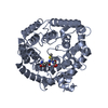 8czlC 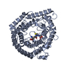 8d0vC 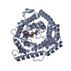 8d19C  3e6uS S: Starting model for refinement C: citing same article ( |
|---|---|
| Similar structure data |
-Links
 PDBj
PDBj


- Assembly
| Deposited unit | 
| ||||||||
|---|---|---|---|---|---|---|---|---|---|
| 1 | 
| ||||||||
| 2 | 
| ||||||||
| Unit cell |
|
-Components
| #1: Protein | Mass: 47504.293 Da / Num. of mol.: 2 Source method: isolated from a genetically manipulated source Source: (gene. exp.) Homo sapiens (human) / Gene: LANCL1, GPR69A / Production host:  Escherichia coli BL21(DE3) (bacteria) / References: UniProt: O43813, glutathione transferase Escherichia coli BL21(DE3) (bacteria) / References: UniProt: O43813, glutathione transferase#2: Protein/peptide | Mass: 710.775 Da / Num. of mol.: 2 / Source method: obtained synthetically Details: The authors report that chain D displays a better fit to electron density for Try 6 than chain C Source: (synth.) synthetic construct (others) #3: Chemical |   Mass: 65.409 Da / Num. of mol.: 2 / Source method: obtained synthetically / Formula: Zn / Feature type: SUBJECT OF INVESTIGATION Mass: 65.409 Da / Num. of mol.: 2 / Source method: obtained synthetically / Formula: Zn / Feature type: SUBJECT OF INVESTIGATION#4: Chemical | Glutathione  Mass: 307.323 Da / Num. of mol.: 2 / Source method: obtained synthetically / Formula: C10H17N3O6S / Feature type: SUBJECT OF INVESTIGATION Mass: 307.323 Da / Num. of mol.: 2 / Source method: obtained synthetically / Formula: C10H17N3O6S / Feature type: SUBJECT OF INVESTIGATION#5: Water | ChemComp-HOH / | Water Mass: 18.015 Da / Num. of mol.: 477 / Source method: isolated from a natural source / Formula: H2O Mass: 18.015 Da / Num. of mol.: 477 / Source method: isolated from a natural source / Formula: H2OHas ligand of interest | Y | |
|---|
-Experimental details
-Experiment
| Experiment | Method: X-RAY DIFFRACTION / Number of used crystals: 1 |
|---|
- Sample preparation
| Crystal | Density Matthews: 2.63 Å3/Da / Density % sol: 53.26 % |
|---|---|
| Crystal grow | Temperature: 282 K / Method: vapor diffusion, hanging drop / pH: 7 Details: 0.70-0.75 M succinic acid pH 7.0, 0.1 M bis-tris-propane pH 7.0 |
-Data collection
| Diffraction | Mean temperature: 100 K / Serial crystal experiment: N |
|---|---|
| Diffraction source | Source: SYNCHROTRON / Site: APS / Beamline: 21-ID-F / Wavelength: 0.97856 Å |
| Detector | Type: RAYONIX MX-300 / Detector: CCD / Date: Feb 11, 2021 |
| Radiation | Protocol: SINGLE WAVELENGTH / Monochromatic (M) / Laue (L): M / Scattering type: x-ray |
| Radiation wavelength | Wavelength: 0.97856 Å / Relative weight: 1 |
| Reflection | Resolution: 1.91→92.5 Å / Num. obs: 75709 / % possible obs: 100 % / Redundancy: 10 % / CC1/2: 0.998 / Rmerge(I) obs: 0.119 / Rpim(I) all: 0.041 / Rrim(I) all: 0.126 / Net I/σ(I): 13.5 |
| Reflection shell | Resolution: 1.914→1.92 Å / Redundancy: 10.1 % / Rmerge(I) obs: 1.284 / Mean I/σ(I) obs: 2.1 / Num. unique obs: 809 / CC1/2: 0.785 / Rpim(I) all: 0.438 / Rrim(I) all: 1.36 / % possible all: 100 |
- Processing
| Software |
| ||||||||||||||||||||||||||||||||||||||||||||||||||||||||||||||||||||||||||||||||||||||||||||||||||||||||||||||||||||||||||||||||||||||||||||||||||||||||||||||||||||||||||||||||||||||
|---|---|---|---|---|---|---|---|---|---|---|---|---|---|---|---|---|---|---|---|---|---|---|---|---|---|---|---|---|---|---|---|---|---|---|---|---|---|---|---|---|---|---|---|---|---|---|---|---|---|---|---|---|---|---|---|---|---|---|---|---|---|---|---|---|---|---|---|---|---|---|---|---|---|---|---|---|---|---|---|---|---|---|---|---|---|---|---|---|---|---|---|---|---|---|---|---|---|---|---|---|---|---|---|---|---|---|---|---|---|---|---|---|---|---|---|---|---|---|---|---|---|---|---|---|---|---|---|---|---|---|---|---|---|---|---|---|---|---|---|---|---|---|---|---|---|---|---|---|---|---|---|---|---|---|---|---|---|---|---|---|---|---|---|---|---|---|---|---|---|---|---|---|---|---|---|---|---|---|---|---|---|---|---|
| Refinement | Method to determine structure: MOLECULAR REPLACEMENT Starting model: 3E6U Resolution: 1.91→25 Å / Cor.coef. Fo:Fc: 0.966 / Cor.coef. Fo:Fc free: 0.955 / SU B: 3.568 / SU ML: 0.099 / Cross valid method: THROUGHOUT / ESU R: 0.145 / ESU R Free: 0.131 / Stereochemistry target values: MAXIMUM LIKELIHOOD / Details: HYDROGENS HAVE BEEN USED IF PRESENT IN THE INPUT
| ||||||||||||||||||||||||||||||||||||||||||||||||||||||||||||||||||||||||||||||||||||||||||||||||||||||||||||||||||||||||||||||||||||||||||||||||||||||||||||||||||||||||||||||||||||||
| Solvent computation | Ion probe radii: 0.8 Å / Shrinkage radii: 0.8 Å / VDW probe radii: 1.2 Å / Solvent model: BABINET MODEL WITH MASK | ||||||||||||||||||||||||||||||||||||||||||||||||||||||||||||||||||||||||||||||||||||||||||||||||||||||||||||||||||||||||||||||||||||||||||||||||||||||||||||||||||||||||||||||||||||||
| Displacement parameters | Biso mean: 31.31 Å2
| ||||||||||||||||||||||||||||||||||||||||||||||||||||||||||||||||||||||||||||||||||||||||||||||||||||||||||||||||||||||||||||||||||||||||||||||||||||||||||||||||||||||||||||||||||||||
| Refinement step | Cycle: 1 / Resolution: 1.91→25 Å
| ||||||||||||||||||||||||||||||||||||||||||||||||||||||||||||||||||||||||||||||||||||||||||||||||||||||||||||||||||||||||||||||||||||||||||||||||||||||||||||||||||||||||||||||||||||||
| Refine LS restraints |
|