 Movie
Movie Controller


+ Open data

- Basic information
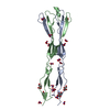
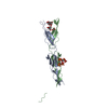
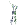
| Entry | Database: PDB / ID: 8ckg | ||||||
|---|---|---|---|---|---|---|---|
| Title | Semaphorin-5A TSR 3-4 domains in complex with sulfate | ||||||
 Components Components | Semaphorin-5A | ||||||
 Keywords Keywords |  SIGNALING PROTEIN / semaphorin / cell signalling / axon guidance cue / glycosaminoglycan binding protein SIGNALING PROTEIN / semaphorin / cell signalling / axon guidance cue / glycosaminoglycan binding protein | ||||||
| Function / homology |  Function and homology information Function and homology informationsignal clustering / diencephalon development / chondroitin sulfate proteoglycan binding / semaphorin receptor binding / positive regulation of actin filament depolymerization / Defective B3GALTL causes PpS / O-glycosylation of TSR domain-containing proteins / Other semaphorin interactions / negative regulation of axon extension involved in axon guidance / syndecan binding ...signal clustering / diencephalon development / chondroitin sulfate proteoglycan binding / semaphorin receptor binding / positive regulation of actin filament depolymerization / Defective B3GALTL causes PpS / O-glycosylation of TSR domain-containing proteins / Other semaphorin interactions / negative regulation of axon extension involved in axon guidance / syndecan binding / blood vessel endothelial cell proliferation involved in sprouting angiogenesis / positive regulation of axon extension involved in axon guidance / chemorepellent activity / negative regulation of cell adhesion / axon extension / heparan sulfate proteoglycan binding / axonal fasciculation / neural crest cell migration / positive regulation of endothelial cell chemotaxis / positive chemotaxis / semaphorin-plexin signaling pathway / negative regulation of endothelial cell apoptotic process / positive regulation of endothelial cell proliferation / cell chemotaxis / axon guidance / positive regulation of angiogenesis / positive regulation of canonical Wnt signaling pathway / cell-cell signaling / nervous system development / positive regulation of phosphatidylinositol 3-kinase/protein kinase B signal transduction / cell adhesion / positive regulation of cell migration / extracellular exosome / membrane / plasma membraneSimilarity search - Function | ||||||
| Biological species |  Homo sapiens (human) Homo sapiens (human) | ||||||
| Method | X-RAY DIFFRACTION / SYNCHROTRON / SAD / Resolution: 1.714 Å | ||||||
 Authors Authors | Nagy, G.N. / Duman, R. / Harlos, K. / El Omari, K. / Wagner, A. / Jones, E.Y. | ||||||
| Funding support |  United Kingdom, 1items United Kingdom, 1items
| ||||||
 Citation Citation | Journal: To Be Published Title: Semaphorin-5A TSR 3-4 domains in complex with sulfate Authors: Nagy, G.N. / Duman, R. / Harlos, K. / El Omari, K. / Wagner, A. / Jones, E.Y. | ||||||
| History |
|
- Structure visualization
| Structure viewer | Molecule: MolmilJmol/JSmol |
|---|
- Downloads & links
-Download
| PDBx/mmCIF format | 8ckg.cif.gz | 104.4 KB | Display | PDBx/mmCIF format |
|---|---|---|---|---|
| PDB format | pdb8ckg.ent.gz | 79.2 KB | Display | PDB format |
| PDBx/mmJSON format | 8ckg.json.gz | Tree view | PDBx/mmJSON format | |
| Others |  Other downloads Other downloads |
-Validation report
| Arichive directory | https://data.pdbj.org/pub/pdb/validation_reports/ck/8ckgftp://data.pdbj.org/pub/pdb/validation_reports/ck/8ckg | HTTPS FTP |
|---|
-Related structure data
| Related structure data | |
|---|---|
| Similar structure data | |
| Experimental dataset #1 | Data reference: 10.6019/EMPIAR-11462 / Data set type: EMPIAR |
-Links
 PDBj
PDBj


- Assembly
| Deposited unit | 
| ||||||||||
|---|---|---|---|---|---|---|---|---|---|---|---|
| 1 |
| ||||||||||
| Unit cell |
| ||||||||||
| Components on special symmetry positions |
|
-Components
| #1: Protein | Mass: 14054.702 Da / Num. of mol.: 2 Source method: isolated from a genetically manipulated source Source: (gene. exp.) Homo sapiens (human) / Gene: SEMA5A, SEMAF / Cell line (production host): HEK293T / Production host: Homo sapiens (human) / References: UniProt: Q13591#2: Chemical | Sulfate  Mass: 96.063 Da / Num. of mol.: 2 / Source method: obtained synthetically / Formula: SO4 / Feature type: SUBJECT OF INVESTIGATION Mass: 96.063 Da / Num. of mol.: 2 / Source method: obtained synthetically / Formula: SO4 / Feature type: SUBJECT OF INVESTIGATION#3: Sugar | ChemComp-MAN / Mannose  Type: D-saccharide, alpha linking / Mass: 180.156 Da / Num. of mol.: 4 / Source method: obtained synthetically / Formula: C6H12O6 / Feature type: SUBJECT OF INVESTIGATION Type: D-saccharide, alpha linking / Mass: 180.156 Da / Num. of mol.: 4 / Source method: obtained synthetically / Formula: C6H12O6 / Feature type: SUBJECT OF INVESTIGATION#4: Water | ChemComp-HOH / | Water Mass: 18.015 Da / Num. of mol.: 99 / Source method: isolated from a natural source / Formula: H2O Mass: 18.015 Da / Num. of mol.: 99 / Source method: isolated from a natural source / Formula: H2OHas ligand of interest | Y | |
|---|
-Experimental details
-Experiment
| Experiment | Method: X-RAY DIFFRACTION / Number of used crystals: 2 |
|---|
- Sample preparation
| Crystal |
| |||||||||||||||
|---|---|---|---|---|---|---|---|---|---|---|---|---|---|---|---|---|
| Crystal grow |
|
-Data collection
| Diffraction |
| |||||||||||||||||||||||||||||||||
|---|---|---|---|---|---|---|---|---|---|---|---|---|---|---|---|---|---|---|---|---|---|---|---|---|---|---|---|---|---|---|---|---|---|---|
| Diffraction source |
| |||||||||||||||||||||||||||||||||
| Detector |
| |||||||||||||||||||||||||||||||||
| Radiation |
| |||||||||||||||||||||||||||||||||
| Radiation wavelength |
| |||||||||||||||||||||||||||||||||
| Reflection | Entry-ID: 8CKG
| |||||||||||||||||||||||||||||||||
| Reflection shell |
|
- Processing
| Software |
| |||||||||||||||||||||||||||||||||||||||||||||||||||||||||||||||||||||||||||||||||||||||||||||||||||||||||||||||||||||||||||||||||||||||||||||||||||||||||||||||||||||||||||||||||||||||||||||||||||||||||||||||||||||||||||||||||
|---|---|---|---|---|---|---|---|---|---|---|---|---|---|---|---|---|---|---|---|---|---|---|---|---|---|---|---|---|---|---|---|---|---|---|---|---|---|---|---|---|---|---|---|---|---|---|---|---|---|---|---|---|---|---|---|---|---|---|---|---|---|---|---|---|---|---|---|---|---|---|---|---|---|---|---|---|---|---|---|---|---|---|---|---|---|---|---|---|---|---|---|---|---|---|---|---|---|---|---|---|---|---|---|---|---|---|---|---|---|---|---|---|---|---|---|---|---|---|---|---|---|---|---|---|---|---|---|---|---|---|---|---|---|---|---|---|---|---|---|---|---|---|---|---|---|---|---|---|---|---|---|---|---|---|---|---|---|---|---|---|---|---|---|---|---|---|---|---|---|---|---|---|---|---|---|---|---|---|---|---|---|---|---|---|---|---|---|---|---|---|---|---|---|---|---|---|---|---|---|---|---|---|---|---|---|---|---|---|---|---|---|---|---|---|---|---|---|---|---|---|---|---|---|---|---|---|
| Refinement | Method to determine structure: SAD / Resolution: 1.714→51.67 Å / SU ML: 0.18 / Cross valid method: FREE R-VALUE / σ(F): 1.35 / Phase error: 30.16 / Stereochemistry target values: ML
| |||||||||||||||||||||||||||||||||||||||||||||||||||||||||||||||||||||||||||||||||||||||||||||||||||||||||||||||||||||||||||||||||||||||||||||||||||||||||||||||||||||||||||||||||||||||||||||||||||||||||||||||||||||||||||||||||
| Solvent computation | Shrinkage radii: 0.9 Å / VDW probe radii: 1.11 Å / Solvent model: FLAT BULK SOLVENT MODEL | |||||||||||||||||||||||||||||||||||||||||||||||||||||||||||||||||||||||||||||||||||||||||||||||||||||||||||||||||||||||||||||||||||||||||||||||||||||||||||||||||||||||||||||||||||||||||||||||||||||||||||||||||||||||||||||||||
| Refinement step | Cycle: LAST / Resolution: 1.714→51.67 Å
| |||||||||||||||||||||||||||||||||||||||||||||||||||||||||||||||||||||||||||||||||||||||||||||||||||||||||||||||||||||||||||||||||||||||||||||||||||||||||||||||||||||||||||||||||||||||||||||||||||||||||||||||||||||||||||||||||
| Refine LS restraints |
| |||||||||||||||||||||||||||||||||||||||||||||||||||||||||||||||||||||||||||||||||||||||||||||||||||||||||||||||||||||||||||||||||||||||||||||||||||||||||||||||||||||||||||||||||||||||||||||||||||||||||||||||||||||||||||||||||
| LS refinement shell |
| |||||||||||||||||||||||||||||||||||||||||||||||||||||||||||||||||||||||||||||||||||||||||||||||||||||||||||||||||||||||||||||||||||||||||||||||||||||||||||||||||||||||||||||||||||||||||||||||||||||||||||||||||||||||||||||||||
| Refinement TLS params. | Method: refined / Refine-ID: X-RAY DIFFRACTION
| |||||||||||||||||||||||||||||||||||||||||||||||||||||||||||||||||||||||||||||||||||||||||||||||||||||||||||||||||||||||||||||||||||||||||||||||||||||||||||||||||||||||||||||||||||||||||||||||||||||||||||||||||||||||||||||||||
| Refinement TLS group |
|
