Journal: PLoS Pathog / Year: 2023 Title: Architecture of the biofilm-associated archaic Chaperone-Usher pilus CupE from Pseudomonas aeruginosa. Authors: Jan Böhning / Adrian W Dobbelstein / Nina Sulkowski / Kira Eilers / Andriko von Kügelgen / Abul K Tarafder / Sew-Yeu Peak-Chew / Mark Skehel / Vikram Alva / Alain Filloux / Tanmay A M Bharat / Abstract: Chaperone-Usher Pathway (CUP) pili are major adhesins in Gram-negative bacteria, mediating bacterial adherence to biotic and abiotic surfaces. While classical CUP pili have been extensively ...Chaperone-Usher Pathway (CUP) pili are major adhesins in Gram-negative bacteria, mediating bacterial adherence to biotic and abiotic surfaces. While classical CUP pili have been extensively characterized, little is known about so-called archaic CUP pili, which are phylogenetically widespread and promote biofilm formation by several human pathogens. In this study, we present the electron cryomicroscopy structure of the archaic CupE pilus from the opportunistic human pathogen Pseudomonas aeruginosa. We show that CupE1 subunits within the pilus are arranged in a zigzag architecture, containing an N-terminal donor β-strand extending from each subunit into the next, where it is anchored by hydrophobic interactions, with comparatively weaker interactions at the rest of the inter-subunit interface. Imaging CupE pili on the surface of P. aeruginosa cells using electron cryotomography shows that CupE pili adopt variable curvatures in response to their environment, which might facilitate their role in promoting cellular attachment. Finally, bioinformatic analysis shows the widespread abundance of cupE genes in isolates of P. aeruginosa and the co-occurrence of cupE with other cup clusters, suggesting interdependence of cup pili in regulating bacterial adherence within biofilms. Taken together, our study provides insights into the architecture of archaic CUP pili, providing a structural basis for understanding their role in promoting cellular adhesion and biofilm formation in P. aeruginosa.
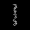
A: SCPU domain-containing protein B: SCPU domain-containing protein C: SCPU domain-containing protein D: SCPU domain-containing protein E: SCPU domain-containing protein
Resolution: 3.5 Å / Resolution method: FSC 0.143 CUT-OFF / Num. of particles: 274457 / Symmetry type: HELICAL
Atomic model building
Protocol: AB INITIO MODEL / Space: REAL Details: Initial local fitting of a homology model was done using ChimeraX, and the structure manually re-built in Coot.
Refine LS restraints
Refine-ID
Type
Dev ideal
Number
ELECTRONMICROSCOPY
f_bond_d
0.002
5740
ELECTRONMICROSCOPY
f_angle_d
0.534
7890
ELECTRONMICROSCOPY
f_dihedral_angle_d
3.641
835
ELECTRONMICROSCOPY
f_chiral_restr
0.048
1005
ELECTRONMICROSCOPY
f_plane_restr
0.003
1015
+
About Yorodumi
-
News
-
Feb 9, 2022. New format data for meta-information of EMDB entries
New format data for meta-information of EMDB entries
Version 3 of the EMDB header file is now the official format.
The previous official version 1.9 will be removed from the archive.
In the structure databanks used in Yorodumi, some data are registered as the other names, "COVID-19 virus" and "2019-nCoV". Here are the details of the virus and the list of structure data.
Jan 31, 2019. EMDB accession codes are about to change! (news from PDBe EMDB page)
EMDB accession codes are about to change! (news from PDBe EMDB page)
The allocation of 4 digits for EMDB accession codes will soon come to an end. Whilst these codes will remain in use, new EMDB accession codes will include an additional digit and will expand incrementally as the available range of codes is exhausted. The current 4-digit format prefixed with “EMD-” (i.e. EMD-XXXX) will advance to a 5-digit format (i.e. EMD-XXXXX), and so on. It is currently estimated that the 4-digit codes will be depleted around Spring 2019, at which point the 5-digit format will come into force.
The EM Navigator/Yorodumi systems omit the EMD- prefix.
Related info.:Q: What is EMD? / ID/Accession-code notation in Yorodumi/EM Navigator
Yorodumi is a browser for structure data from EMDB, PDB, SASBDB, etc.
This page is also the successor to EM Navigator detail page, and also detail information page/front-end page for Omokage search.
The word "yorodu" (or yorozu) is an old Japanese word meaning "ten thousand". "mi" (miru) is to see.
Related info.:EMDB / PDB / SASBDB / Comparison of 3 databanks / Yorodumi Search / Aug 31, 2016. New EM Navigator & Yorodumi / Yorodumi Papers / Jmol/JSmol / Function and homology information / Changes in new EM Navigator and Yorodumi
 Movie
Movie Controller


 Open data
 Basic information
 Components
 Keywords
 Pseudomonas aeruginosa /
  Function and homology information
 Authors
 United Kingdom,
  United States, 7items
  Citation
 Structure visualization
 Downloads & links
 Other downloads
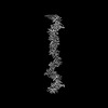
 PDBj
PDBj Assembly
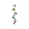
 Sample preparation
 Electron microscopy imaging
 Processing