 Movie
Movie Controller

[English] 日本語
 Yorodumi
- PDB-8caq: Structure of Tau filaments Type I from Subacute Sclerosing Panenc... -
+ Open data

- Basic information
| Entry | Database: PDB / ID: 8caq | ||||||
|---|---|---|---|---|---|---|---|
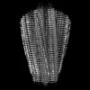
| Title | Structure of Tau filaments Type I from Subacute Sclerosing Panencephalitis | ||||||
 Components Components | Microtubule-associated protein tau Tau protein Tau protein | ||||||
 Keywords Keywords | PROTEIN FIBRIL / Tau filament / SSPE / Neurodegenerative disease | ||||||
| Function / homology |  Function and homology information Function and homology informationplus-end-directed organelle transport along microtubule / axonal transport / histone-dependent DNA binding / neurofibrillary tangle assembly / positive regulation of diacylglycerol kinase activity / negative regulation of establishment of protein localization to mitochondrion / neurofibrillary tangle / positive regulation of protein localization to synapse / microtubule lateral binding / tubulin complex ...plus-end-directed organelle transport along microtubule / axonal transport / histone-dependent DNA binding / neurofibrillary tangle assembly / positive regulation of diacylglycerol kinase activity / negative regulation of establishment of protein localization to mitochondrion / neurofibrillary tangle / positive regulation of protein localization to synapse / microtubule lateral binding / tubulin complex / phosphatidylinositol bisphosphate binding / main axon / regulation of long-term synaptic depression / negative regulation of kinase activity / negative regulation of tubulin deacetylation / generation of neurons / regulation of chromosome organization / positive regulation of protein localization / rRNA metabolic process / internal protein amino acid acetylation / regulation of mitochondrial fission / lipoprotein particle binding / intracellular distribution of mitochondria / axonal transport of mitochondrion / axon development / central nervous system neuron development / regulation of microtubule polymerization / microtubule polymerization / minor groove of adenine-thymine-rich DNA binding / negative regulation of mitochondrial membrane potential / dynactin binding / glial cell projection / apolipoprotein binding / protein polymerization / negative regulation of mitochondrial fission / axolemma / Caspase-mediated cleavage of cytoskeletal proteins / regulation of microtubule polymerization or depolymerization / positive regulation of axon extension / supramolecular fiber organization / Activation of AMPK downstream of NMDARs / regulation of microtubule cytoskeleton organization / stress granule assembly / cytoplasmic microtubule organization / regulation of cellular response to heat / regulation of calcium-mediated signaling / axon cytoplasm / positive regulation of microtubule polymerization / cellular response to brain-derived neurotrophic factor stimulus / somatodendritic compartment / synapse assembly / phosphatidylinositol binding / nuclear periphery / cellular response to nerve growth factor stimulus / positive regulation of superoxide anion generation / protein phosphatase 2A binding / regulation of autophagy / astrocyte activation / synapse organization / microglial cell activation / response to lead ion / Hsp90 protein binding / regulation of synaptic plasticity / PKR-mediated signaling / protein homooligomerization / cytoplasmic ribonucleoprotein granule / memory / microtubule cytoskeleton organization / cellular response to reactive oxygen species / SH3 domain binding / neuron projection development / activation of cysteine-type endopeptidase activity involved in apoptotic process / microtubule cytoskeleton / protein-macromolecule adaptor activity / single-stranded DNA binding / cell-cell signaling / cellular response to heat / cell body / actin binding / growth cone / protein-folding chaperone binding / double-stranded DNA binding / microtubule binding / microtubule / amyloid fibril formation / sequence-specific DNA binding / dendritic spine / learning or memory / neuron projection / nuclear speck / membrane raft / axon / negative regulation of gene expression / dendrite / neuronal cell body / DNA damage response / protein kinase binding / enzyme binding / mitochondrion / DNA bindingSimilarity search - Function | ||||||
| Biological species |  Homo sapiens (human) Homo sapiens (human) | ||||||
| Method | ELECTRON MICROSCOPY / helical reconstruction / cryo EM / Resolution: 2.3 Å | ||||||
 Authors Authors | Qi, C. / Hasegawa, M. / Takao, M. / Sakai, M. / Akagi, M. / Iwasaki, Y. / Yoshida, M. / Scheres, S.H.W. / Goedert, M. | ||||||
| Funding support |  United Kingdom, 1items United Kingdom, 1items
| ||||||
 Citation Citation | Journal: Acta Neuropathol Commun / Year: 2023 Title: Identical tau filaments in subacute sclerosing panencephalitis and chronic traumatic encephalopathy. Authors: Chao Qi / Masato Hasegawa / Masaki Takao / Motoko Sakai / Mayasuki Sasaki / Masashi Mizutani / Akio Akagi / Yasushi Iwasaki / Hiroaki Miyahara / Mari Yoshida / Sjors H W Scheres / Michel Goedert /  Abstract: Subacute sclerosing panencephalitis (SSPE) occurs in some individuals after measles infection, following a symptom-free period of several years. It resembles chronic traumatic encephalopathy (CTE), ...Subacute sclerosing panencephalitis (SSPE) occurs in some individuals after measles infection, following a symptom-free period of several years. It resembles chronic traumatic encephalopathy (CTE), which happens after repetitive head impacts or exposure to blast waves, following a symptom-free period. As in CTE, the neurofibrillary changes of SSPE are concentrated in superficial cortical layers. Here we used electron cryo-microscopy (cryo-EM) of tau filaments from two cases of SSPE to show that the tau folds of SSPE and CTE are identical. Two types of filaments were each made of two identical protofilaments with an extra density in the β-helix region. Like in CTE, the vast majority of tau filaments were Type I, with a minority of Type II filaments. These findings suggest that the CTE tau fold can be caused by different environmental insults, which may be linked by inflammatory changes. | ||||||
| History |
|
- Structure visualization
| Structure viewer | Molecule: MolmilJmol/JSmol |
|---|
- Downloads & links
-Download
| PDBx/mmCIF format | 8caq.cif.gz | 103.8 KB | Display | PDBx/mmCIF format |
|---|---|---|---|---|
| PDB format | pdb8caq.ent.gz | 67.6 KB | Display | PDB format |
| PDBx/mmJSON format | 8caq.json.gz | Tree view | PDBx/mmJSON format | |
| Others |  Other downloads Other downloads |
-Validation report
| Arichive directory | https://data.pdbj.org/pub/pdb/validation_reports/ca/8caqftp://data.pdbj.org/pub/pdb/validation_reports/ca/8caq | HTTPS FTP |
|---|
-Related structure data
| Related structure data |  16532MC 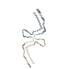 8caxC M: map data used to model this data C: citing same article ( |
|---|---|
| Similar structure data |
-Links
 PDBj
PDBj


- Assembly
| Deposited unit | 
|
|---|---|
| 1 |
|
-Components
| #1: Protein | Tau protein / Neurofibrillary tangle protein / Paired helical filament-tau / PHF-tau Mass: 45919.871 Da / Num. of mol.: 5 / Source method: isolated from a natural source / Source: (natural) Homo sapiens (human) / References: UniProt: P10636 |
|---|
-Experimental details
-Experiment
| Experiment | Method: ELECTRON MICROSCOPY |
|---|---|
| EM experiment | Aggregation state: FILAMENT / 3D reconstruction method: helical reconstruction |
- Sample preparation
| Component | Name: tau / Type: COMPLEX / Entity ID: all / Source: NATURAL |
|---|---|
| Source (natural) | Organism: Homo sapiens (human) |
| Buffer solution | pH: 7.5 |
| Specimen | Embedding applied: NO / Shadowing applied: NO / Staining applied: NO / Vitrification applied: YES |
| Vitrification | Cryogen name: ETHANE |
- Electron microscopy imaging
| Experimental equipment |  Model: Titan Krios / Image courtesy: FEI Company |
|---|---|
| Microscopy | Model: FEI TITAN KRIOS |
| Electron gun | Electron source: FIELD EMISSION GUN / Accelerating voltage: 300 kV / Illumination mode: FLOOD BEAM |
| Electron lens | Mode: BRIGHT FIELDBright-field microscopy / Nominal defocus max: 2000 nm / Nominal defocus min: 1000 nm |
| Image recording | Electron dose: 40 e/Å2 / Film or detector model: FEI FALCON IV (4k x 4k) |
- Processing
| CTF correction | Type: NONE |
|---|---|
| Helical symmerty | Angular rotation/subunit: 179.35 ° / Axial rise/subunit: 2.38 Å / Axial symmetry: C1 |
| 3D reconstruction | Resolution: 2.3 Å / Resolution method: FSC 0.143 CUT-OFF / Num. of particles: 222257 / Symmetry type: HELICAL |
