 Movie
Movie Controller

[English] 日本語
 Yorodumi
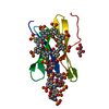
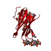
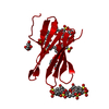
- PDB-8c9z: The RSL - sulfonato-calix[8]arene complex, H32 form, citrate pH 6.0 -
+ Open data

- Basic information
| Entry | Database: PDB / ID: 8c9z | ||||||||||||
|---|---|---|---|---|---|---|---|---|---|---|---|---|---|
| Title | The RSL - sulfonato-calix[8]arene complex, H32 form, citrate pH 6.0 | ||||||||||||
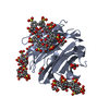
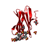
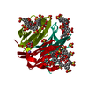
 Components Components | Fucose-binding lectin protein | ||||||||||||
 Keywords Keywords | SUGAR BINDING PROTEIN /  Macrocycle / Calixarene / Lectin Macrocycle / Calixarene / Lectin | ||||||||||||
| Function / homology | Fucose-specific lectin / Fungal fucose-specific lectin / carbohydrate binding / beta-D-fructopyranose / sulfonato-calix[8]arene / Fucose-binding lectin protein Function and homology information Function and homology information | ||||||||||||
| Biological species |  Ralstonia solanacearum (bacteria) Ralstonia solanacearum (bacteria) | ||||||||||||
| Method | X-RAY DIFFRACTION / SYNCHROTRON / MOLECULAR REPLACEMENT / Resolution: 1.18 Å | ||||||||||||
 Authors Authors | Mockler, N.M. / Ramberg, K. / Crowley, P.B. | ||||||||||||
| Funding support |  Ireland, 3items Ireland, 3items
| ||||||||||||
 Citation Citation | Journal: Acta Crystallogr D Struct Biol / Year: 2023 Title: Protein-macrocycle polymorphism: crystal form IV of the Ralstonia solanacearum lectin-sulfonato-calix[8]arene complex. Authors: Mockler, N.M. / Ramberg, K.O. / Crowley, P.B. | ||||||||||||
| History |
|
- Structure visualization
| Structure viewer | Molecule: MolmilJmol/JSmol |
|---|
- Downloads & links
-Download
| PDBx/mmCIF format | 8c9z.cif.gz | 41.6 KB | Display | PDBx/mmCIF format |
|---|---|---|---|---|
| PDB format | pdb8c9z.ent.gz | 27 KB | Display | PDB format |
| PDBx/mmJSON format | 8c9z.json.gz | Tree view | PDBx/mmJSON format | |
| Others |  Other downloads Other downloads |
-Validation report
| Arichive directory | https://data.pdbj.org/pub/pdb/validation_reports/c9/8c9zftp://data.pdbj.org/pub/pdb/validation_reports/c9/8c9z | HTTPS FTP |
|---|
-Related structure data
-Links
 PDBj
PDBj- Assembly
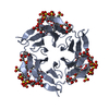
| Deposited unit | 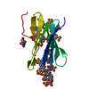
| |||||||||||||||||||||||||||
|---|---|---|---|---|---|---|---|---|---|---|---|---|---|---|---|---|---|---|---|---|---|---|---|---|---|---|---|---|
| 1 |
| |||||||||||||||||||||||||||
| Unit cell |
| |||||||||||||||||||||||||||
| Components on special symmetry positions |
|
-Components
| #1: Protein | Mass: 9733.562 Da / Num. of mol.: 1 Source method: isolated from a genetically manipulated source Source: (gene. exp.) Ralstonia solanacearum (bacteria)Gene: E7Z57_08365, RSP795_21825, RSP822_19650, RUN39_v1_50103 Production host: Escherichia coli (E. coli) / References: UniProt: A0A0S4TLR1 | ||||||||
|---|---|---|---|---|---|---|---|---|---|
| #2: Sugar | Fructose  Type: D-saccharide, beta linking / Mass: 180.156 Da / Num. of mol.: 2 / Source method: obtained synthetically / Formula: C6H12O6 Type: D-saccharide, beta linking / Mass: 180.156 Da / Num. of mol.: 2 / Source method: obtained synthetically / Formula: C6H12O6#3: Chemical | 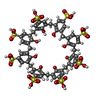  Mass: 1489.481 Da / Num. of mol.: 2 / Source method: obtained synthetically / Formula: C56H48O32S8 / Feature type: SUBJECT OF INVESTIGATION Mass: 1489.481 Da / Num. of mol.: 2 / Source method: obtained synthetically / Formula: C56H48O32S8 / Feature type: SUBJECT OF INVESTIGATION#4: Chemical | Glycerol  Mass: 92.094 Da / Num. of mol.: 2 / Source method: isolated from a natural source / Formula: C3H8O3 Mass: 92.094 Da / Num. of mol.: 2 / Source method: isolated from a natural source / Formula: C3H8O3#5: Water | ChemComp-HOH / | Water Mass: 18.015 Da / Num. of mol.: 103 / Source method: isolated from a natural source / Formula: H2O Mass: 18.015 Da / Num. of mol.: 103 / Source method: isolated from a natural source / Formula: H2OHas ligand of interest | Y | |
-Experimental details
-Experiment
| Experiment | Method: X-RAY DIFFRACTION / Number of used crystals: 1 |
|---|
- Sample preparation
| Crystal | Density Matthews: 2.49 Å3/Da / Density % sol: 51 % |
|---|---|
| Crystal grow | Temperature: 293 K / Method: vapor diffusion, hanging drop / pH: 6 / Details: 1.2 M tri-sodium citrate pH 6.0 |
-Data collection
| Diffraction | Mean temperature: 100 K / Serial crystal experiment: N |
|---|---|
| Diffraction source | Source: SYNCHROTRON / Site: SOLEIL  / Beamline: PROXIMA 2 / Wavelength: 0.98 Å / Beamline: PROXIMA 2 / Wavelength: 0.98 Å |
| Detector | Type: DECTRIS EIGER X 9M / Detector: PIXEL / Date: Jul 9, 2022 |
| Radiation | Monochromator: M / Protocol: SINGLE WAVELENGTH / Monochromatic (M) / Laue (L): M / Scattering type: x-ray |
| Radiation wavelength | Wavelength: 0.98 Å / Relative weight: 1 |
| Reflection | Resolution: 1.18→57.02 Å / Num. obs: 41706 / % possible obs: 99.8 % / Redundancy: 17.3 % / Biso Wilson estimate: 13.3 Å2 / CC1/2: 1 / Rmerge(I) obs: 0.065 / Rpim(I) all: 0.016 / Rrim(I) all: 0.067 / Net I/σ(I): 19 |
| Reflection shell | Resolution: 1.18→1.2 Å / Redundancy: 8.9 % / Rmerge(I) obs: 1.304 / Mean I/σ(I) obs: 1.8 / Num. unique obs: 1963 / CC1/2: 0.622 / Rpim(I) all: 0.448 / % possible all: 96.3 |
- Processing
| Software |
| ||||||||||||||||||||||||||||||||||||||||||||||||||||||||||||||||||||||||||||||||||||||||||||||||||||||||||||||||
|---|---|---|---|---|---|---|---|---|---|---|---|---|---|---|---|---|---|---|---|---|---|---|---|---|---|---|---|---|---|---|---|---|---|---|---|---|---|---|---|---|---|---|---|---|---|---|---|---|---|---|---|---|---|---|---|---|---|---|---|---|---|---|---|---|---|---|---|---|---|---|---|---|---|---|---|---|---|---|---|---|---|---|---|---|---|---|---|---|---|---|---|---|---|---|---|---|---|---|---|---|---|---|---|---|---|---|---|---|---|---|---|---|---|
| Refinement | Method to determine structure: MOLECULAR REPLACEMENT / Resolution: 1.18→43.04 Å / SU ML: 0.1027 / Cross valid method: FREE R-VALUE / σ(F): 1.34 / Phase error: 19.3653 Stereochemistry target values: GeoStd + Monomer Library + CDL v1.2
| ||||||||||||||||||||||||||||||||||||||||||||||||||||||||||||||||||||||||||||||||||||||||||||||||||||||||||||||||
| Solvent computation | Shrinkage radii: 0.9 Å / VDW probe radii: 1.11 Å / Solvent model: FLAT BULK SOLVENT MODEL | ||||||||||||||||||||||||||||||||||||||||||||||||||||||||||||||||||||||||||||||||||||||||||||||||||||||||||||||||
| Displacement parameters | Biso mean: 18.93 Å2 | ||||||||||||||||||||||||||||||||||||||||||||||||||||||||||||||||||||||||||||||||||||||||||||||||||||||||||||||||
| Refinement step | Cycle: LAST / Resolution: 1.18→43.04 Å
| ||||||||||||||||||||||||||||||||||||||||||||||||||||||||||||||||||||||||||||||||||||||||||||||||||||||||||||||||
| Refine LS restraints |
| ||||||||||||||||||||||||||||||||||||||||||||||||||||||||||||||||||||||||||||||||||||||||||||||||||||||||||||||||
| LS refinement shell |
|
