 Movie
Movie Controller


+ Open data

- Basic information
| Entry | Database: PDB / ID: 8c7p | ||||||
|---|---|---|---|---|---|---|---|
| Title | Tagless BtuM in complex with cyanocobalamin | ||||||
 Components Components | Cobalamin ABC transporter | ||||||
 Keywords Keywords |  TRANSPORT PROTEIN / Cobalamin / Membrane transporter TRANSPORT PROTEIN / Cobalamin / Membrane transporter | ||||||
| Function / homology | membrane => GO:0016020 / COBALAMIN / Uncharacterized protein Function and homology information Function and homology information | ||||||
| Biological species |  Thiobacillus denitrificans ATCC 25259 (bacteria) Thiobacillus denitrificans ATCC 25259 (bacteria) | ||||||
| Method | X-RAY DIFFRACTION / SYNCHROTRON / MOLECULAR REPLACEMENT / Resolution: 3 Å | ||||||
 Authors Authors | Martinez-Felices, J.M. / Slotboom, D.J. | ||||||
| Funding support |  Netherlands, 1items Netherlands, 1items
| ||||||
 Citation Citation | Journal: To Be Published Title: Structure of the Thiobacillus denitrificans cobalamin transporter BtuM in complex with cyanocobalamin Authors: Martinez-Felices, J.M. / Slotboom, D.J. | ||||||
| History |
|
- Structure visualization
| Structure viewer | Molecule: MolmilJmol/JSmol |
|---|
- Downloads & links
-Download
| PDBx/mmCIF format | 8c7p.cif.gz | 111.1 KB | Display | PDBx/mmCIF format |
|---|---|---|---|---|
| PDB format | pdb8c7p.ent.gz | 65.4 KB | Display | PDB format |
| PDBx/mmJSON format | 8c7p.json.gz | Tree view | PDBx/mmJSON format | |
| Others |  Other downloads Other downloads |
-Validation report
| Arichive directory | https://data.pdbj.org/pub/pdb/validation_reports/c7/8c7pftp://data.pdbj.org/pub/pdb/validation_reports/c7/8c7p | HTTPS FTP |
|---|
-Related structure data
| Related structure data | 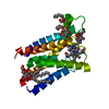 8ovvC  6ffv C: citing same article ( |
|---|---|
| Similar structure data |
-Links
 PDBj
PDBj- Assembly
| Deposited unit | 
| ||||||||
|---|---|---|---|---|---|---|---|---|---|
| 1 |
| ||||||||
| Unit cell |
|
-Components
| #1: Protein | Mass: 21794.711 Da / Num. of mol.: 1 Source method: isolated from a genetically manipulated source Details: Histidine tag and a portion of the 3C cleavage tag are cleaved during the purification prior to crystallization. Source: (gene. exp.) Thiobacillus denitrificans ATCC 25259 (bacteria)Gene: Tbd_2719 / Plasmid: pBAD24 Details (production host): Ampicilin resistance, L-Arabinose induced polymerase Production host: Escherichia coli MC1061 (bacteria) / References: UniProt: Q3SFD8 |
|---|---|
| #2: Chemical | ChemComp-B12 / Vitamin B12  Mass: 1330.356 Da / Num. of mol.: 1 / Source method: obtained synthetically / Formula: C62H89CoN13O14P / Feature type: SUBJECT OF INVESTIGATION Mass: 1330.356 Da / Num. of mol.: 1 / Source method: obtained synthetically / Formula: C62H89CoN13O14P / Feature type: SUBJECT OF INVESTIGATION |
| Has ligand of interest | Y |
-Experimental details
-Experiment
| Experiment | Method: X-RAY DIFFRACTION / Number of used crystals: 1 |
|---|
- Sample preparation
| Crystal | Density Matthews: 2.14 Å3/Da / Density % sol: 42.53 % / Description: Coloured bars |
|---|---|
| Crystal grow | Temperature: 277 K / Method: vapor diffusion, sitting drop / pH: 5.5 Details: 0.2 M Potassium chloride, 0.1 M Sodium citrate pH 5.5, 37 % v/v Pentaerythritol propoxylate (PO/OH). |
-Data collection
| Diffraction | Mean temperature: 100 K / Serial crystal experiment: N |
|---|---|
| Diffraction source | Source: SYNCHROTRON / Site: Diamond  / Beamline: I24 / Wavelength: 0.976 Å / Beamline: I24 / Wavelength: 0.976 Å |
| Detector | Type: DECTRIS PILATUS3 6M / Detector: PIXEL / Date: Jun 15, 2020 |
| Radiation | Protocol: SINGLE WAVELENGTH / Monochromatic (M) / Laue (L): M / Scattering type: x-ray |
| Radiation wavelength | Wavelength: 0.976 Å / Relative weight: 1 |
| Reflection | Resolution: 2.528→79.084 Å / Num. obs: 6814 / % possible obs: 99.9 % / Redundancy: 2.21 % / Biso Wilson estimate: 49.73 Å2 / CC1/2: 0.98 / Net I/σ(I): 10.4 |
| Reflection shell | Resolution: 2.53→2.63 Å / Redundancy: 0.9 % / Mean I/σ(I) obs: 1.5 / Num. unique obs: 43 / CC1/2: 0.53 / % possible all: 2.8 |
- Processing
| Software |
| |||||||||||||||||||||||||||||||||||||||||||||||||||||||||||||||||||||||||||||||||||||||||||||||||||||||||||||||||||||||||||||||||||||||||||||||||||||||||||||||||||||||||||||||||||||||||||||||||||||||||||||||||||||||||||||||||||||||
|---|---|---|---|---|---|---|---|---|---|---|---|---|---|---|---|---|---|---|---|---|---|---|---|---|---|---|---|---|---|---|---|---|---|---|---|---|---|---|---|---|---|---|---|---|---|---|---|---|---|---|---|---|---|---|---|---|---|---|---|---|---|---|---|---|---|---|---|---|---|---|---|---|---|---|---|---|---|---|---|---|---|---|---|---|---|---|---|---|---|---|---|---|---|---|---|---|---|---|---|---|---|---|---|---|---|---|---|---|---|---|---|---|---|---|---|---|---|---|---|---|---|---|---|---|---|---|---|---|---|---|---|---|---|---|---|---|---|---|---|---|---|---|---|---|---|---|---|---|---|---|---|---|---|---|---|---|---|---|---|---|---|---|---|---|---|---|---|---|---|---|---|---|---|---|---|---|---|---|---|---|---|---|---|---|---|---|---|---|---|---|---|---|---|---|---|---|---|---|---|---|---|---|---|---|---|---|---|---|---|---|---|---|---|---|---|---|---|---|---|---|---|---|---|---|---|---|---|---|---|---|---|---|
| Refinement | Method to determine structure: MOLECULAR REPLACEMENT / Resolution: 3→79.084 Å / Cor.coef. Fo:Fc: 0.907 / Cor.coef. Fo:Fc free: 0.841 / WRfactor Rfree: 0.264 / WRfactor Rwork: 0.216 / SU B: 33.707 / SU ML: 0.275 / Average fsc free: 0.9556 / Average fsc work: 0.9658 / Cross valid method: FREE R-VALUE / ESU R: 0.719 / ESU R Free: 0.428 / Details: Hydrogens have not been used
| |||||||||||||||||||||||||||||||||||||||||||||||||||||||||||||||||||||||||||||||||||||||||||||||||||||||||||||||||||||||||||||||||||||||||||||||||||||||||||||||||||||||||||||||||||||||||||||||||||||||||||||||||||||||||||||||||||||||
| Solvent computation | Ion probe radii: 0.8 Å / Shrinkage radii: 0.8 Å / VDW probe radii: 1.2 Å / Solvent model: BABINET MODEL PLUS MASK | |||||||||||||||||||||||||||||||||||||||||||||||||||||||||||||||||||||||||||||||||||||||||||||||||||||||||||||||||||||||||||||||||||||||||||||||||||||||||||||||||||||||||||||||||||||||||||||||||||||||||||||||||||||||||||||||||||||||
| Displacement parameters | Biso mean: 81.507 Å2
| |||||||||||||||||||||||||||||||||||||||||||||||||||||||||||||||||||||||||||||||||||||||||||||||||||||||||||||||||||||||||||||||||||||||||||||||||||||||||||||||||||||||||||||||||||||||||||||||||||||||||||||||||||||||||||||||||||||||
| Refinement step | Cycle: LAST / Resolution: 3→79.084 Å
| |||||||||||||||||||||||||||||||||||||||||||||||||||||||||||||||||||||||||||||||||||||||||||||||||||||||||||||||||||||||||||||||||||||||||||||||||||||||||||||||||||||||||||||||||||||||||||||||||||||||||||||||||||||||||||||||||||||||
| Refine LS restraints |
| |||||||||||||||||||||||||||||||||||||||||||||||||||||||||||||||||||||||||||||||||||||||||||||||||||||||||||||||||||||||||||||||||||||||||||||||||||||||||||||||||||||||||||||||||||||||||||||||||||||||||||||||||||||||||||||||||||||||
| LS refinement shell | Refine-ID: X-RAY DIFFRACTION / Total num. of bins used: 20
| |||||||||||||||||||||||||||||||||||||||||||||||||||||||||||||||||||||||||||||||||||||||||||||||||||||||||||||||||||||||||||||||||||||||||||||||||||||||||||||||||||||||||||||||||||||||||||||||||||||||||||||||||||||||||||||||||||||||
| Refinement TLS params. | Method: refined / Origin x: -41.9798 Å / Origin y: 19.713 Å / Origin z: 7.2118 Å
| |||||||||||||||||||||||||||||||||||||||||||||||||||||||||||||||||||||||||||||||||||||||||||||||||||||||||||||||||||||||||||||||||||||||||||||||||||||||||||||||||||||||||||||||||||||||||||||||||||||||||||||||||||||||||||||||||||||||
| Refinement TLS group | Selection: ALL |
