 Movie
Movie Controller


+ Open data

- Basic information
| Entry | Database: PDB / ID: 8c6f | ||||||
|---|---|---|---|---|---|---|---|
| Title | Light SFX structure of D.m(6-4)photolyase at 400fs time delay | ||||||
 Components Components | Cryptochrome-1 | ||||||
 Keywords Keywords | FLAVOPROTEIN / photolyase / SFX / FAD | ||||||
| Function / homology |  Function and homology informationdeoxyribodipyrimidine photo-lyase activity / entrainment of circadian clock by photoperiod / FAD binding / circadian regulation of gene expression / DNA binding / nucleus / cytoplasm Function and homology informationdeoxyribodipyrimidine photo-lyase activity / entrainment of circadian clock by photoperiod / FAD binding / circadian regulation of gene expression / DNA binding / nucleus / cytoplasmSimilarity search - Function | ||||||
| Biological species |  Drosophila melanogaster (fruit fly) Drosophila melanogaster (fruit fly) | ||||||
| Method | X-RAY DIFFRACTION / FREE ELECTRON LASER / MOLECULAR REPLACEMENT / Resolution: 1.9 Å | ||||||
 Authors Authors | Cellini, A. / Kumar, M. / Nimmrich, A. / Mutisya, J. / Furrer, A. / Beale, E.V. / Carrillo, M. / Malla, T.N. / Maj, P. / Dworkowskic, F. ...Cellini, A. / Kumar, M. / Nimmrich, A. / Mutisya, J. / Furrer, A. / Beale, E.V. / Carrillo, M. / Malla, T.N. / Maj, P. / Dworkowskic, F. / Cirelli, C. / Ozerovi, D. / Bacellar, C. / Strandfuss, J. / Weinert, T. / Ihalainen, J.A. / Yuan Wahlgren, W. / Westenhoff, S. | ||||||
| Funding support | European Union, 1items
| ||||||
 Citation Citation | Journal: Nat.Chem. / Year: 2024 Title: Directed ultrafast conformational changes accompany electron transfer in a photolyase as resolved by serial crystallography. Authors: Cellini, A. / Shankar, M.K. / Nimmrich, A. / Hunt, L.A. / Monrroy, L. / Mutisya, J. / Furrer, A. / Beale, E.V. / Carrillo, M. / Malla, T.N. / Maj, P. / Vrhovac, L. / Dworkowski, F. / ...Authors: Cellini, A. / Shankar, M.K. / Nimmrich, A. / Hunt, L.A. / Monrroy, L. / Mutisya, J. / Furrer, A. / Beale, E.V. / Carrillo, M. / Malla, T.N. / Maj, P. / Vrhovac, L. / Dworkowski, F. / Cirelli, C. / Johnson, P.J.M. / Ozerov, D. / Stojkovic, E.A. / Hammarstrom, L. / Bacellar, C. / Standfuss, J. / Maj, M. / Schmidt, M. / Weinert, T. / Ihalainen, J.A. / Wahlgren, W.Y. / Westenhoff, S. | ||||||
| History |
|
- Structure visualization
| Structure viewer | Molecule: MolmilJmol/JSmol |
|---|
- Downloads & links
-Download
| PDBx/mmCIF format | 8c6f.cif.gz | 156.3 KB | Display | PDBx/mmCIF format |
|---|---|---|---|---|
| PDB format | pdb8c6f.ent.gz | 97.3 KB | Display | PDB format |
| PDBx/mmJSON format | 8c6f.json.gz | Tree view | PDBx/mmJSON format | |
| Others |  Other downloads Other downloads |
-Validation report
| Arichive directory | https://data.pdbj.org/pub/pdb/validation_reports/c6/8c6fftp://data.pdbj.org/pub/pdb/validation_reports/c6/8c6f | HTTPS FTP |
|---|
-Related structure data
| Related structure data | 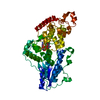 8c1uC  8c69C 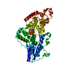 8c6aC  8c6bC 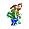 8c6cC  8c6hC C: citing same article ( |
|---|---|
| Similar structure data |
-Links
 PDBj
PDBj
- Assembly
| Deposited unit | 
| ||||||||||||
|---|---|---|---|---|---|---|---|---|---|---|---|---|---|
| 1 |
| ||||||||||||
| Unit cell |
|
-Components
| #1: Protein | Mass: 58079.805 Da / Num. of mol.: 1 Source method: isolated from a genetically manipulated source Source: (gene. exp.) Drosophila melanogaster (fruit fly) / Gene: phr6-4, CG2488 / Production host:  Escherichia coli (E. coli) / References: UniProt: Q8SXK5 Escherichia coli (E. coli) / References: UniProt: Q8SXK5 |
|---|---|
| #2: Chemical | ChemComp-FAD / Flavin adenine dinucleotide  Mass: 785.550 Da / Num. of mol.: 1 / Source method: obtained synthetically / Formula: C27H33N9O15P2 / Feature type: SUBJECT OF INVESTIGATION / Comment: FAD*YM Mass: 785.550 Da / Num. of mol.: 1 / Source method: obtained synthetically / Formula: C27H33N9O15P2 / Feature type: SUBJECT OF INVESTIGATION / Comment: FAD*YM |
| #3: Chemical | ChemComp-GOL / Glycerol  Mass: 92.094 Da / Num. of mol.: 1 / Source method: obtained synthetically / Formula: C3H8O3 Mass: 92.094 Da / Num. of mol.: 1 / Source method: obtained synthetically / Formula: C3H8O3 |
| #4: Water | ChemComp-HOH / Water Mass: 18.015 Da / Num. of mol.: 134 / Source method: isolated from a natural source / Formula: H2O Mass: 18.015 Da / Num. of mol.: 134 / Source method: isolated from a natural source / Formula: H2O |
| Has ligand of interest | Y |
-Experimental details
-Experiment
| Experiment | Method: X-RAY DIFFRACTION / Number of used crystals: 1 |
|---|
- Sample preparation
| Crystal | Density Matthews: 2.43 Å3/Da / Density % sol: 49.28 % |
|---|---|
| Crystal grow | Temperature: 277.15 K / Method: batch mode / pH: 6.5 Details: 100 mM bis-tris pH=6.5, 200 mM lithium sulphate monohydrate, 22 % PEG 3350, 0.5 % Ethyl acetate. |
-Data collection
| Diffraction | Mean temperature: 293.15 K / Serial crystal experiment: Y |
|---|---|
| Diffraction source | Source: FREE ELECTRON LASER / Site: SwissFEL ARAMIS  / Beamline: ESA / Wavelength: 1 Å / Beamline: ESA / Wavelength: 1 Å |
| Detector | Type: PSI JUNGFRAU 16M / Detector: PIXEL / Date: Sep 27, 2021 |
| Radiation | Protocol: SINGLE WAVELENGTH / Monochromatic (M) / Laue (L): M / Scattering type: x-ray |
| Radiation wavelength | Wavelength: 1 Å / Relative weight: 1 |
| Reflection | Resolution: 1.9→15.72 Å / Num. obs: 44136 / % possible obs: 99.99 % / Redundancy: 56.25 % / Biso Wilson estimate: 3.4 Å2 / R split: 0.15 / Net I/σ(I): 6.53 |
| Reflection shell | Resolution: 1.9→1.91 Å / Num. unique obs: 1764 / R split: 0.51 |
| Serial crystallography sample delivery | Method: injection |
- Processing
| Software |
| ||||||||||||||||||||||||||||||||||||||||||||||||||||||||||||||||||||||||||||||||||||||||||||||||||||||||||||||||
|---|---|---|---|---|---|---|---|---|---|---|---|---|---|---|---|---|---|---|---|---|---|---|---|---|---|---|---|---|---|---|---|---|---|---|---|---|---|---|---|---|---|---|---|---|---|---|---|---|---|---|---|---|---|---|---|---|---|---|---|---|---|---|---|---|---|---|---|---|---|---|---|---|---|---|---|---|---|---|---|---|---|---|---|---|---|---|---|---|---|---|---|---|---|---|---|---|---|---|---|---|---|---|---|---|---|---|---|---|---|---|---|---|---|
| Refinement | Method to determine structure: MOLECULAR REPLACEMENT / Resolution: 1.9→15.72 Å / SU ML: 0.3292 / Cross valid method: FREE R-VALUE / σ(F): 0 / Phase error: 33.367 Stereochemistry target values: GeoStd + Monomer Library + CDL v1.2
| ||||||||||||||||||||||||||||||||||||||||||||||||||||||||||||||||||||||||||||||||||||||||||||||||||||||||||||||||
| Solvent computation | Shrinkage radii: 0.9 Å / VDW probe radii: 1.11 Å / Solvent model: FLAT BULK SOLVENT MODEL | ||||||||||||||||||||||||||||||||||||||||||||||||||||||||||||||||||||||||||||||||||||||||||||||||||||||||||||||||
| Displacement parameters | Biso mean: 20.75 Å2 | ||||||||||||||||||||||||||||||||||||||||||||||||||||||||||||||||||||||||||||||||||||||||||||||||||||||||||||||||
| Refinement step | Cycle: LAST / Resolution: 1.9→15.72 Å
| ||||||||||||||||||||||||||||||||||||||||||||||||||||||||||||||||||||||||||||||||||||||||||||||||||||||||||||||||
| Refine LS restraints |
| ||||||||||||||||||||||||||||||||||||||||||||||||||||||||||||||||||||||||||||||||||||||||||||||||||||||||||||||||
| LS refinement shell |
| ||||||||||||||||||||||||||||||||||||||||||||||||||||||||||||||||||||||||||||||||||||||||||||||||||||||||||||||||
| Refinement TLS params. | Method: refined / Refine-ID: X-RAY DIFFRACTION
| ||||||||||||||||||||||||||||||||||||||||||||||||||||||||||||||||||||||||||||||||||||||||||||||||||||||||||||||||
| Refinement TLS group | Refine-ID: X-RAY DIFFRACTION / Auth asym-ID: A / Label asym-ID: A
|
