 Movie
Movie Controller


+ Open data

- Basic information
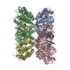
| Entry | Database: PDB / ID: 8c2z | |||||||||
|---|---|---|---|---|---|---|---|---|---|---|
| Title | Crystal structure of DYRK1B in complex with AZ191 | |||||||||
 Components Components | Dual specificity tyrosine-phosphorylation-regulated kinase 1B | |||||||||
 Keywords Keywords |  TRANSFERASE / Kinase / diabetes / kinase inhibitor TRANSFERASE / Kinase / diabetes / kinase inhibitor | |||||||||
| Function / homology |  Function and homology information Function and homology informationmyoblast fusion / dual-specificity kinase / adipose tissue development / protein serine/threonine/tyrosine kinase activity / chromosome / protein tyrosine kinase activity / transcription coactivator activity / protein kinase activity / protein phosphorylation / protein serine kinase activity ...myoblast fusion / dual-specificity kinase / adipose tissue development / protein serine/threonine/tyrosine kinase activity / chromosome / protein tyrosine kinase activity / transcription coactivator activity / protein kinase activity / protein phosphorylation / protein serine kinase activity / DNA repair / protein serine/threonine kinase activity / nucleolus / positive regulation of DNA-templated transcription / nucleoplasm / ATP binding / nucleusSimilarity search - Function | |||||||||
| Biological species |  Homo sapiens (human) Homo sapiens (human) | |||||||||
| Method | X-RAY DIFFRACTION / SYNCHROTRON / MOLECULAR REPLACEMENT / Resolution: 1.91 Å | |||||||||
 Authors Authors | Grygier, P. / Pustelny, K. / Dubin, G. / Czarna, A. | |||||||||
| Funding support |  Poland, 2items Poland, 2items
| |||||||||
 Citation Citation | Journal: To Be Published Title: Structural perspective on the design of selective DYRK1B inhibitors Authors: Grygier, P. / Pustelny, K. / Dubin, G. / Czarna, A. | |||||||||
| History |
|
- Structure visualization
| Structure viewer | Molecule: MolmilJmol/JSmol |
|---|
- Downloads & links
-Download
| PDBx/mmCIF format | 8c2z.cif.gz | 174.3 KB | Display | PDBx/mmCIF format |
|---|---|---|---|---|
| PDB format | pdb8c2z.ent.gz | 119.8 KB | Display | PDB format |
| PDBx/mmJSON format | 8c2z.json.gz | Tree view | PDBx/mmJSON format | |
| Others |  Other downloads Other downloads |
-Validation report
| Arichive directory | https://data.pdbj.org/pub/pdb/validation_reports/c2/8c2zftp://data.pdbj.org/pub/pdb/validation_reports/c2/8c2z | HTTPS FTP |
|---|
-Related structure data
-Links
 PDBj
PDBj- Assembly
| Deposited unit | 
| ||||||||||
|---|---|---|---|---|---|---|---|---|---|---|---|
| 1 |
| ||||||||||
| Unit cell |
|
-Components
| #1: Protein | Mass: 42904.121 Da / Num. of mol.: 1 Source method: isolated from a genetically manipulated source Source: (gene. exp.) Homo sapiens (human) / Gene: DYRK1B, MIRK / Production host:  Escherichia coli (E. coli) / References: UniProt: Q9Y463, dual-specificity kinase Escherichia coli (E. coli) / References: UniProt: Q9Y463, dual-specificity kinase | ||||
|---|---|---|---|---|---|
| #2: Chemical | ChemComp-QS0 /   Mass: 429.517 Da / Num. of mol.: 1 / Source method: isolated from a natural source / Formula: C24H27N7O / Feature type: SUBJECT OF INVESTIGATION Mass: 429.517 Da / Num. of mol.: 1 / Source method: isolated from a natural source / Formula: C24H27N7O / Feature type: SUBJECT OF INVESTIGATION | ||||
| #3: Chemical |   Mass: 54.938 Da / Num. of mol.: 3 / Source method: isolated from a natural source / Formula: Mn Mass: 54.938 Da / Num. of mol.: 3 / Source method: isolated from a natural source / Formula: Mn#4: Water | ChemComp-HOH / | Water Mass: 18.015 Da / Num. of mol.: 185 / Source method: isolated from a natural source / Formula: H2O Mass: 18.015 Da / Num. of mol.: 185 / Source method: isolated from a natural source / Formula: H2OHas ligand of interest | Y | |
-Experimental details
-Experiment
| Experiment | Method: X-RAY DIFFRACTION / Number of used crystals: 1 |
|---|
- Sample preparation
| Crystal | Density Matthews: 2.22 Å3/Da / Density % sol: 44.5 % |
|---|---|
| Crystal grow | Temperature: 293.15 K / Method: vapor diffusion, sitting drop Details: 0.1 M Bis-Tris pH 5.5, 25% PEG3350, 0.2 M magnesium chloride, 0.1 M manganese (II) chloride. |
-Data collection
| Diffraction | Mean temperature: 100 K / Serial crystal experiment: N |
|---|---|
| Diffraction source | Source: SYNCHROTRON / Site: PETRA III, DESY  / Beamline: P11 / Wavelength: 1.0332 Å / Beamline: P11 / Wavelength: 1.0332 Å |
| Detector | Type: DECTRIS EIGER2 X 16M / Detector: PIXEL / Date: Aug 15, 2022 |
| Radiation | Protocol: SINGLE WAVELENGTH / Monochromatic (M) / Laue (L): M / Scattering type: x-ray |
| Radiation wavelength | Wavelength: 1.0332 Å / Relative weight: 1 |
| Reflection | Resolution: 1.91→45.41 Å / Num. obs: 29064 / % possible obs: 97.1 % / Redundancy: 8.3 % / Biso Wilson estimate: 38.16 Å2 / CC1/2: 0.999 / Rmerge(I) obs: 0.078 / Rpim(I) all: 0.029 / Rrim(I) all: 0.083 / Χ2: 1.04 / Net I/σ(I): 13.8 / Num. measured all: 242154 |
| Reflection shell | Resolution: 1.91→1.95 Å / % possible obs: 97.8 % / Redundancy: 8.5 % / Rmerge(I) obs: 2.035 / Num. measured all: 16608 / Num. unique obs: 1945 / CC1/2: 0.453 / Rpim(I) all: 0.726 / Rrim(I) all: 2.164 / Χ2: 1.08 / Net I/σ(I) obs: 1.3 |
- Processing
| Software |
| ||||||||||||||||||||||||||||||||||||||||||||||||||||||||||||||||||||||||||||||||||||||||||||||||||||
|---|---|---|---|---|---|---|---|---|---|---|---|---|---|---|---|---|---|---|---|---|---|---|---|---|---|---|---|---|---|---|---|---|---|---|---|---|---|---|---|---|---|---|---|---|---|---|---|---|---|---|---|---|---|---|---|---|---|---|---|---|---|---|---|---|---|---|---|---|---|---|---|---|---|---|---|---|---|---|---|---|---|---|---|---|---|---|---|---|---|---|---|---|---|---|---|---|---|---|---|---|---|
| Refinement | Method to determine structure: MOLECULAR REPLACEMENT / Resolution: 1.91→45.41 Å / SU ML: 0.2807 / Cross valid method: FREE R-VALUE / σ(F): 1.34 / Phase error: 24.7947 Stereochemistry target values: GeoStd + Monomer Library + CDL v1.2
| ||||||||||||||||||||||||||||||||||||||||||||||||||||||||||||||||||||||||||||||||||||||||||||||||||||
| Solvent computation | Shrinkage radii: 0.9 Å / VDW probe radii: 1.1 Å / Solvent model: FLAT BULK SOLVENT MODEL | ||||||||||||||||||||||||||||||||||||||||||||||||||||||||||||||||||||||||||||||||||||||||||||||||||||
| Displacement parameters | Biso mean: 49.03 Å2 | ||||||||||||||||||||||||||||||||||||||||||||||||||||||||||||||||||||||||||||||||||||||||||||||||||||
| Refinement step | Cycle: LAST / Resolution: 1.91→45.41 Å
| ||||||||||||||||||||||||||||||||||||||||||||||||||||||||||||||||||||||||||||||||||||||||||||||||||||
| Refine LS restraints |
| ||||||||||||||||||||||||||||||||||||||||||||||||||||||||||||||||||||||||||||||||||||||||||||||||||||
| LS refinement shell |
| ||||||||||||||||||||||||||||||||||||||||||||||||||||||||||||||||||||||||||||||||||||||||||||||||||||
| Refinement TLS params. | Method: refined / Refine-ID: X-RAY DIFFRACTION
| ||||||||||||||||||||||||||||||||||||||||||||||||||||||||||||||||||||||||||||||||||||||||||||||||||||
| Refinement TLS group | Refine-ID: X-RAY DIFFRACTION / Auth asym-ID: A / Label asym-ID: A
|
