 Movie
Movie Controller


+ Open data

- Basic information
| Entry | Database: PDB / ID: 8btj | ||||||
|---|---|---|---|---|---|---|---|
| Title | Murine cytomegalovirus protein M35 | ||||||
 Components Components | Protein M35 | ||||||
 Keywords Keywords |  STRUCTURAL PROTEIN / CMV / M35 / virus protein / NFKB mediated transcription STRUCTURAL PROTEIN / CMV / M35 / virus protein / NFKB mediated transcription | ||||||
| Function / homology | Herpesvirus phosphoprotein 85 (HHV6-7 U14/HCMV UL25) / Herpesvirus phosphoprotein 85 (HHV6-7 U14/HCMV UL25) / host cell nucleus / (R,R)-2,3-BUTANEDIOL / MALONATE ION / Protein M35 Function and homology information Function and homology information | ||||||
| Biological species |  Muromegalovirus Muromegalovirus | ||||||
| Method | X-RAY DIFFRACTION / SYNCHROTRON / MOLECULAR REPLACEMENT / Resolution: 1.94 Å | ||||||
 Authors Authors | Schmelz, S. / Van den Heuvel, J. / Blankenfeldt, W. | ||||||
| Funding support | 1items
| ||||||
 Citation Citation | Journal: J.Virol. / Year: 2023 Title: The Cytomegalovirus M35 Protein Directly Binds to the Interferon-beta Enhancer and Modulates Transcription of Ifnb1 and Other IRF3-Driven Genes. Authors: Schwanke, H. / Goncalves Magalhaes, V. / Schmelz, S. / Wyler, E. / Hennig, T. / Gunther, T. / Grundhoff, A. / Dolken, L. / Landthaler, M. / van Ham, M. / Jansch, L. / Bussow, K. / van den ...Authors: Schwanke, H. / Goncalves Magalhaes, V. / Schmelz, S. / Wyler, E. / Hennig, T. / Gunther, T. / Grundhoff, A. / Dolken, L. / Landthaler, M. / van Ham, M. / Jansch, L. / Bussow, K. / van den Heuvel, J. / Blankenfeldt, W. / Friedel, C.C. / Erhard, F. / Brinkmann, M.M. | ||||||
| History |
|
- Structure visualization
| Structure viewer | Molecule: MolmilJmol/JSmol |
|---|
- Downloads & links
-Download
| PDBx/mmCIF format | 8btj.cif.gz | 395.8 KB | Display | PDBx/mmCIF format |
|---|---|---|---|---|
| PDB format | pdb8btj.ent.gz | 263.4 KB | Display | PDB format |
| PDBx/mmJSON format | 8btj.json.gz | Tree view | PDBx/mmJSON format | |
| Others |  Other downloads Other downloads |
-Validation report
| Arichive directory | https://data.pdbj.org/pub/pdb/validation_reports/bt/8btjftp://data.pdbj.org/pub/pdb/validation_reports/bt/8btj | HTTPS FTP |
|---|
-Related structure data
| Similar structure data |
|---|
-Links
 PDBj
PDBj- Assembly
| Deposited unit | 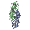
| ||||||||||||
|---|---|---|---|---|---|---|---|---|---|---|---|---|---|
| 1 |
| ||||||||||||
| Unit cell |
|
-Components
| #1: Protein | Mass: 50834.816 Da / Num. of mol.: 2 Source method: isolated from a genetically manipulated source Source: (gene. exp.) Muromegalovirus / Gene: M35 / Cell line (production host): High Five / Production host:  Trichoplusia ni (cabbage looper) / References: UniProt: A8E1G1 Trichoplusia ni (cabbage looper) / References: UniProt: A8E1G1#2: Chemical | ChemComp-BU3 / (   Mass: 90.121 Da / Num. of mol.: 10 / Source method: obtained synthetically / Formula: C4H10O2 Mass: 90.121 Da / Num. of mol.: 10 / Source method: obtained synthetically / Formula: C4H10O2#3: Chemical | ChemComp-MLI / | Malonic acid  Mass: 102.046 Da / Num. of mol.: 1 / Source method: obtained synthetically / Formula: C3H2O4 Mass: 102.046 Da / Num. of mol.: 1 / Source method: obtained synthetically / Formula: C3H2O4#4: Water | ChemComp-HOH / | Water Mass: 18.015 Da / Num. of mol.: 299 / Source method: isolated from a natural source / Formula: H2O Mass: 18.015 Da / Num. of mol.: 299 / Source method: isolated from a natural source / Formula: H2OHas ligand of interest | N | |
|---|
-Experimental details
-Experiment
| Experiment | Method: X-RAY DIFFRACTION / Number of used crystals: 1 |
|---|
- Sample preparation
| Crystal | Density Matthews: 2.52 Å3/Da / Density % sol: 51.2 % |
|---|---|
| Crystal grow | Temperature: 293 K / Method: vapor diffusion, sitting drop Details: 1.1M Na2 Malon, 0.1 M HEPES pH 7.0, 0.5 % (v/v) Jeffamine ED-2001 protein conc: 3.6mg/ml cryoprotectant: 12 (v/v) (2R,3R) -(-)-2,3-Butanediol |
-Data collection
| Diffraction | Mean temperature: 100 K / Serial crystal experiment: N |
|---|---|
| Diffraction source | Source: SYNCHROTRON / Site: PETRA III, DESY  / Beamline: P11 / Wavelength: 0.978 Å / Beamline: P11 / Wavelength: 0.978 Å |
| Detector | Type: DECTRIS PILATUS 6M-F / Detector: PIXEL / Date: Sep 12, 2017 |
| Radiation | Protocol: SINGLE WAVELENGTH / Monochromatic (M) / Laue (L): M / Scattering type: x-ray |
| Radiation wavelength | Wavelength: 0.978 Å / Relative weight: 1 |
| Reflection | Resolution: 1.94→66.52 Å / Num. obs: 50243 / % possible obs: 92 % / Redundancy: 10.6 % / Biso Wilson estimate: 29.79 Å2 / CC1/2: 0.999 / Rpim(I) all: 0.047 / Net I/σ(I): 11.6 |
| Reflection shell | Resolution: 1.94→2.151 Å / Num. unique obs: 2513 / CC1/2: 0.581 / Rpim(I) all: 0.488 |
- Processing
| Software |
| |||||||||||||||||||||||||||||||||||||||||||||||||||||||||||||||||||||||||||||||||||||||||||||||||||||||||||||||||||||||||||||||||||||
|---|---|---|---|---|---|---|---|---|---|---|---|---|---|---|---|---|---|---|---|---|---|---|---|---|---|---|---|---|---|---|---|---|---|---|---|---|---|---|---|---|---|---|---|---|---|---|---|---|---|---|---|---|---|---|---|---|---|---|---|---|---|---|---|---|---|---|---|---|---|---|---|---|---|---|---|---|---|---|---|---|---|---|---|---|---|---|---|---|---|---|---|---|---|---|---|---|---|---|---|---|---|---|---|---|---|---|---|---|---|---|---|---|---|---|---|---|---|---|---|---|---|---|---|---|---|---|---|---|---|---|---|---|---|---|
| Refinement | Method to determine structure: MOLECULAR REPLACEMENT / Resolution: 1.94→57.95 Å / SU ML: 0.2236 / Cross valid method: FREE R-VALUE / σ(F): 1.35 / Phase error: 28.2547 Stereochemistry target values: GeoStd + Monomer Library + CDL v1.2
| |||||||||||||||||||||||||||||||||||||||||||||||||||||||||||||||||||||||||||||||||||||||||||||||||||||||||||||||||||||||||||||||||||||
| Solvent computation | Shrinkage radii: 0.9 Å / VDW probe radii: 1.1 Å / Solvent model: FLAT BULK SOLVENT MODEL | |||||||||||||||||||||||||||||||||||||||||||||||||||||||||||||||||||||||||||||||||||||||||||||||||||||||||||||||||||||||||||||||||||||
| Displacement parameters | Biso mean: 36.96 Å2 | |||||||||||||||||||||||||||||||||||||||||||||||||||||||||||||||||||||||||||||||||||||||||||||||||||||||||||||||||||||||||||||||||||||
| Refinement step | Cycle: LAST / Resolution: 1.94→57.95 Å
| |||||||||||||||||||||||||||||||||||||||||||||||||||||||||||||||||||||||||||||||||||||||||||||||||||||||||||||||||||||||||||||||||||||
| Refine LS restraints |
| |||||||||||||||||||||||||||||||||||||||||||||||||||||||||||||||||||||||||||||||||||||||||||||||||||||||||||||||||||||||||||||||||||||
| LS refinement shell |
|
