 Movie
Movie Controller


+ Open data

- Basic information
| Entry | Database: PDB / ID: 8bpf | |||||||||||||||||||||
|---|---|---|---|---|---|---|---|---|---|---|---|---|---|---|---|---|---|---|---|---|---|---|
| Title | FcMR binding at subunit Fcu1 of IgM pentamer | |||||||||||||||||||||
 Components Components |
| |||||||||||||||||||||
 Keywords Keywords |  IMMUNE SYSTEM / IgM Fc receptor IMMUNE SYSTEM / IgM Fc receptor | |||||||||||||||||||||
| Function / homology |  Function and homology information Function and homology informationhigh-affinity IgM receptor activity / immunoglobulin transcytosis in epithelial cells / IgM binding / regulation of B cell receptor signaling pathway / polymeric immunoglobulin binding / Fc receptor-mediated immune complex endocytosis / humoral immune response mediated by circulating immunoglobulin / cellular defense response / trans-Golgi network membrane / early endosome membrane ...high-affinity IgM receptor activity / immunoglobulin transcytosis in epithelial cells / IgM binding / regulation of B cell receptor signaling pathway / polymeric immunoglobulin binding / Fc receptor-mediated immune complex endocytosis / humoral immune response mediated by circulating immunoglobulin / cellular defense response / trans-Golgi network membrane / early endosome membrane / lysosomal membrane / negative regulation of apoptotic process / extracellular region / plasma membraneSimilarity search - Function | |||||||||||||||||||||
| Biological species |  Homo sapiens (human) Homo sapiens (human) | |||||||||||||||||||||
| Method | ELECTRON MICROSCOPY / single particle reconstruction / cryo EM / Resolution: 3.5 Å | |||||||||||||||||||||
 Authors Authors | Chen, Q. / Rosenthal, P. / Tolar, P. | |||||||||||||||||||||
| Funding support |  United Kingdom, 6items United Kingdom, 6items
| |||||||||||||||||||||
 Citation Citation | Journal: Nat Struct Mol Biol / Year: 2023 Title: Structural basis for Fc receptor recognition of immunoglobulin M. Authors: Qu Chen / Rajesh P Menon / Laura Masino / Pavel Tolar / Peter B Rosenthal / Abstract: Immunoglobulin Fc receptors are cell surface transmembrane proteins that bind to the Fc constant region of antibodies and play critical roles in regulating immune responses by activation of immune ...Immunoglobulin Fc receptors are cell surface transmembrane proteins that bind to the Fc constant region of antibodies and play critical roles in regulating immune responses by activation of immune cells, clearance of immune complexes and regulation of antibody production. FcμR is the immunoglobulin M (IgM) antibody isotype-specific Fc receptor involved in the survival and activation of B cells. Here we reveal eight binding sites for the human FcμR immunoglobulin domain on the IgM pentamer by cryogenic electron microscopy. One of the sites overlaps with the binding site for the polymeric immunoglobulin receptor (pIgR), but a different mode of FcμR binding explains its antibody isotype specificity. Variation in FcμR binding sites and their occupancy reflects the asymmetry of the IgM pentameric core and the versatility of FcμR binding. The complex explains engagement with polymeric serum IgM and the monomeric IgM B-cell receptor (BCR). | |||||||||||||||||||||
| History |
|
- Structure visualization
| Structure viewer | Molecule: MolmilJmol/JSmol |
|---|
- Downloads & links
-Download
| PDBx/mmCIF format | 8bpf.cif.gz | 423.7 KB | Display | PDBx/mmCIF format |
|---|---|---|---|---|
| PDB format | pdb8bpf.ent.gz | 344.8 KB | Display | PDB format |
| PDBx/mmJSON format | 8bpf.json.gz | Tree view | PDBx/mmJSON format | |
| Others |  Other downloads Other downloads |
-Validation report
| Arichive directory | https://data.pdbj.org/pub/pdb/validation_reports/bp/8bpfftp://data.pdbj.org/pub/pdb/validation_reports/bp/8bpf | HTTPS FTP |
|---|
-Related structure data
| Related structure data |  16151MC 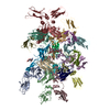 8bpeC 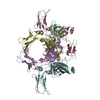 8bpgC M: map data used to model this data C: citing same article ( |
|---|---|
| Similar structure data |
-Links
 PDBj
PDBj- Assembly
| Deposited unit | 
|
|---|---|
| 1 |
|
-Components
| #1: Protein | Mass: 38343.086 Da / Num. of mol.: 10 / Source method: isolated from a natural source / Source: (natural) Homo sapiens (human)#2: Protein | | Mass: 26020.617 Da / Num. of mol.: 1 Source method: isolated from a genetically manipulated source Source: (gene. exp.) Homo sapiens (human) / Gene: FCMR, FAIM3, TOSO / Cell line (production host): HEK293 / Production host: Homo sapiens (human) / References: UniProt: O60667#3: Protein | | Mass: 18120.586 Da / Num. of mol.: 1 / Source method: isolated from a natural source / Source: (natural) Homo sapiens (human)#4: Polysaccharide | 2-acetamido-2-deoxy-beta-D-glucopyranose-(1-4)-2-acetamido-2-deoxy-beta-D-glucopyranose | / Mass: 424.401 Da / Num. of mol.: 1Source method: isolated from a genetically manipulated source #5: Sugar | ChemComp-NAG / N-Acetylglucosamine  Type: D-saccharide, beta linking / Mass: 221.208 Da / Num. of mol.: 10 / Source method: obtained synthetically / Formula: C8H15NO6 / Feature type: SUBJECT OF INVESTIGATION Type: D-saccharide, beta linking / Mass: 221.208 Da / Num. of mol.: 10 / Source method: obtained synthetically / Formula: C8H15NO6 / Feature type: SUBJECT OF INVESTIGATIONHas ligand of interest | Y | Sequence details | The entities of this entry have been subject to varying amounts of proteolysis resulting in ...The entities of this entry have been subject to varying amounts of proteolysis resulting in different sequence lengths for the same protein. | |
|---|
-Experimental details
-Experiment
| Experiment | Method: ELECTRON MICROSCOPY |
|---|---|
| EM experiment | Aggregation state: PARTICLE / 3D reconstruction method: single particle reconstruction |
- Sample preparation
| Component | Name: human FcMR/IgM-Fc complex / Type: COMPLEX / Entity ID: #1, #3, #2 / Source: RECOMBINANT |
|---|---|
| Source (natural) | Organism: Homo sapiens (human) |
| Source (recombinant) | Organism: Homo sapiens (human) |
| Buffer solution | pH: 7.5 |
| Specimen | Conc.: 0.1 mg/ml / Embedding applied: NO / Shadowing applied: NO / Staining applied: NO / Vitrification applied: YES |
| Vitrification | Cryogen name: ETHANE |
- Electron microscopy imaging
| Experimental equipment |  Model: Titan Krios / Image courtesy: FEI Company |
|---|---|
| Microscopy | Model: FEI TITAN KRIOS |
| Electron gun | Electron source: FIELD EMISSION GUN / Accelerating voltage: 300 kV / Illumination mode: FLOOD BEAM |
| Electron lens | Mode: BRIGHT FIELDBright-field microscopy / Nominal defocus max: 3500 nm / Nominal defocus min: 1200 nm / Calibrated defocus min: 1000 nm / Calibrated defocus max: 4000 nm / Cs: 2.7 mm / C2 aperture diameter: 50 µm |
| Specimen holder | Cryogen: NITROGEN / Specimen holder model: FEI TITAN KRIOS AUTOGRID HOLDER |
| Image recording | Electron dose: 50.6 e/Å2 / Detector mode: COUNTING / Film or detector model: GATAN K2 QUANTUM (4k x 4k) |
- Processing
| Software | Name: PHENIX / Version: 1.19.2_4158: / Classification: refinement | ||||||||||||||||||||||||
|---|---|---|---|---|---|---|---|---|---|---|---|---|---|---|---|---|---|---|---|---|---|---|---|---|---|
| CTF correction | Type: PHASE FLIPPING AND AMPLITUDE CORRECTION | ||||||||||||||||||||||||
| 3D reconstruction | Resolution: 3.5 Å / Resolution method: FSC 0.143 CUT-OFF / Num. of particles: 539010 / Symmetry type: POINT | ||||||||||||||||||||||||
| Atomic model building | Space: REAL | ||||||||||||||||||||||||
| Refine LS restraints |
|
