 Movie
Movie Controller


+ Open data

- Basic information
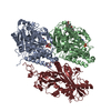
| Entry | Database: PDB / ID: 8bjs | |||||||||
|---|---|---|---|---|---|---|---|---|---|---|
| Title | Apo KIF20A[55-510] crystal structure | |||||||||
 Components Components |
| |||||||||
 Keywords Keywords |  MOTOR PROTEIN / Kinesin 6 / motor domain / ATPase / mitosis / cytokinesis / Golgi / microtubule MOTOR PROTEIN / Kinesin 6 / motor domain / ATPase / mitosis / cytokinesis / Golgi / microtubule | |||||||||
| Function / homology |  Function and homology information Function and homology informationMitotic Telophase/Cytokinesis / Kinesins / COPI-dependent Golgi-to-ER retrograde traffic / midbody abscission / MHC class II antigen presentation / microtubule bundle formation / kinesin complex / microtubule motor activity / intercellular bridge / microtubule-based movement ...Mitotic Telophase/Cytokinesis / Kinesins / COPI-dependent Golgi-to-ER retrograde traffic / midbody abscission / MHC class II antigen presentation / microtubule bundle formation / kinesin complex / microtubule motor activity / intercellular bridge / microtubule-based movement / mitotic cytokinesis / regulation of cytokinesis / spindle / protein transport / midbody / microtubule binding / microtubule / protein kinase binding / Golgi apparatus / ATP hydrolysis activity / nucleoplasm / ATP bindingSimilarity search - Function | |||||||||
| Biological species |  Mus musculus (house mouse) Mus musculus (house mouse) | |||||||||
| Method | X-RAY DIFFRACTION / SYNCHROTRON / MOLECULAR REPLACEMENT / Resolution: 2.73 Å | |||||||||
 Authors Authors | Ranaivoson, F.M. / Kikuti, C. / Crozet, V. / Sirkia, M.E. / Houdusse, A. | |||||||||
| Funding support |  France, 2items France, 2items
| |||||||||
 Citation Citation | Journal: Open Biol / Year: 2023 Title: Nucleotide-free structures of KIF20A illuminate atypical mechanochemistry in this kinesin-6. Authors: Fanomezana Moutse Ranaivoson / Vincent Crozet / Matthieu P M H Benoit / Amna Abdalla Mohammed Khalid / Carlos Kikuti / Helena Sirkia / Ahmed El Marjou / Stéphanie Miserey-Lenkei / Ana B ...Authors: Fanomezana Moutse Ranaivoson / Vincent Crozet / Matthieu P M H Benoit / Amna Abdalla Mohammed Khalid / Carlos Kikuti / Helena Sirkia / Ahmed El Marjou / Stéphanie Miserey-Lenkei / Ana B Asenjo / Hernando Sosa / Christoph F Schmidt / Steven S Rosenfeld / Anne Houdusse /   Abstract: KIF20A is a critical kinesin for cell division and a promising anti-cancer drug target. The mechanisms underlying its cellular roles remain elusive. Interestingly, unusual coupling between the ...KIF20A is a critical kinesin for cell division and a promising anti-cancer drug target. The mechanisms underlying its cellular roles remain elusive. Interestingly, unusual coupling between the nucleotide- and microtubule-binding sites of this kinesin-6 has been reported, but little is known about how its divergent sequence leads to atypical motility properties. We present here the first high-resolution structure of its motor domain that delineates the highly unusual structural features of this motor, including a long L6 insertion that integrates into the core of the motor domain and that drastically affects allostery and ATPase activity. Together with the high-resolution cryo-electron microscopy microtubule-bound KIF20A structure that reveals the microtubule-binding interface, we dissect the peculiarities of the KIF20A sequence that influence its mechanochemistry, leading to low motility compared to other kinesins. Structural and functional insights from the KIF20A pre-power stroke conformation highlight the role of extended insertions in shaping the motor's mechanochemical cycle. Essential for force production and processivity is the length of the neck linker in kinesins. We highlight here the role of the sequence preceding the neck linker in controlling its backward docking and show that a neck linker four times longer than that in kinesin-1 is required for the activity of this motor. | |||||||||
| History |
|
- Structure visualization
| Structure viewer | Molecule: MolmilJmol/JSmol |
|---|
- Downloads & links
-Download
| PDBx/mmCIF format | 8bjs.cif.gz | 159.3 KB | Display | PDBx/mmCIF format |
|---|---|---|---|---|
| PDB format | pdb8bjs.ent.gz | 121.5 KB | Display | PDB format |
| PDBx/mmJSON format | 8bjs.json.gz | Tree view | PDBx/mmJSON format | |
| Others |  Other downloads Other downloads |
-Validation report
| Arichive directory | https://data.pdbj.org/pub/pdb/validation_reports/bj/8bjsftp://data.pdbj.org/pub/pdb/validation_reports/bj/8bjs | HTTPS FTP |
|---|
-Related structure data


-Links
 PDBj
PDBj


- Assembly
| Deposited unit | 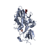
| ||||||||
|---|---|---|---|---|---|---|---|---|---|
| 1 |
| ||||||||
| Unit cell |
|
-Components
| #1: Protein | KIF20A / Kinesin-like protein 174 / Rab6-interacting kinesin-like protein / Rabkinesin-6 Mass: 52450.281 Da / Num. of mol.: 1 Source method: isolated from a genetically manipulated source Source: (gene. exp.) Mus musculus (house mouse) / Gene: Kif20a, Rab6kifl / Production host:  Escherichia coli (E. coli) / References: UniProt: P97329 Escherichia coli (E. coli) / References: UniProt: P97329 |
|---|---|
| #2: Protein/peptide | Mass: 358.434 Da / Num. of mol.: 1 Source method: isolated from a genetically manipulated source Source: (gene. exp.) Mus musculus (house mouse) / Production host: Escherichia coli (E. coli) |
| #3: Chemical | ChemComp-SO4 / Sulfate  Mass: 96.063 Da / Num. of mol.: 1 / Source method: obtained synthetically / Formula: SO4 Mass: 96.063 Da / Num. of mol.: 1 / Source method: obtained synthetically / Formula: SO4 |
| #4: Water | ChemComp-HOH / Water Mass: 18.015 Da / Num. of mol.: 46 / Source method: isolated from a natural source / Formula: H2O Mass: 18.015 Da / Num. of mol.: 46 / Source method: isolated from a natural source / Formula: H2O |
| Has ligand of interest | N |
-Experimental details
-Experiment
| Experiment | Method: X-RAY DIFFRACTION / Number of used crystals: 1 |
|---|
- Sample preparation
| Crystal | Density Matthews: 2.15 Å3/Da / Density % sol: 42.87 % |
|---|---|
| Crystal grow | Temperature: 293 K / Method: vapor diffusion, hanging drop Details: 1.8 M NaCl, 0.5 M (NH4)2SO4, 0.1 M Bis-Tris, pH 5.5 |
-Data collection
| Diffraction | Mean temperature: 100 K / Serial crystal experiment: N |
|---|---|
| Diffraction source | Source: SYNCHROTRON / Site: SOLEIL / Beamline: PROXIMA 1 / Wavelength: 0.979 Å |
| Detector | Type: DECTRIS PILATUS 6M / Detector: PIXEL / Date: Jan 26, 2018 |
| Radiation | Protocol: SINGLE WAVELENGTH / Monochromatic (M) / Laue (L): M / Scattering type: x-ray |
| Radiation wavelength | Wavelength: 0.979 Å / Relative weight: 1 |
| Reflection | Resolution: 2.73→64.24 Å / Num. obs: 12169 / % possible obs: 99.85 % / Redundancy: 6.9 % / CC1/2: 0.995 / Rmerge(I) obs: 0.1332 / Rrim(I) all: 0.1441 / Net I/σ(I): 9.5 |
| Reflection shell | Resolution: 2.73→2.83 Å / Rmerge(I) obs: 0.6778 / Mean I/σ(I) obs: 2.35 / Num. unique obs: 1209 / CC1/2: 0.887 / Rrim(I) all: 0.7307 |
- Processing
| Software |
| ||||||||||||||||||||||||||||||||||||||||||||||||||||||||||||||||||||||||||||||||||||||||||||||||||||||||||||
|---|---|---|---|---|---|---|---|---|---|---|---|---|---|---|---|---|---|---|---|---|---|---|---|---|---|---|---|---|---|---|---|---|---|---|---|---|---|---|---|---|---|---|---|---|---|---|---|---|---|---|---|---|---|---|---|---|---|---|---|---|---|---|---|---|---|---|---|---|---|---|---|---|---|---|---|---|---|---|---|---|---|---|---|---|---|---|---|---|---|---|---|---|---|---|---|---|---|---|---|---|---|---|---|---|---|---|---|---|---|
| Refinement | Method to determine structure: MOLECULAR REPLACEMENT Starting model: Se-SAD Structure Resolution: 2.73→54.63 Å / Cor.coef. Fo:Fc: 0.917 / Cor.coef. Fo:Fc free: 0.897 / SU R Cruickshank DPI: 0.95 / Cross valid method: THROUGHOUT / σ(F): 0 / SU R Blow DPI: 0.868 / SU Rfree Blow DPI: 0.31 / SU Rfree Cruickshank DPI: 0.317
| ||||||||||||||||||||||||||||||||||||||||||||||||||||||||||||||||||||||||||||||||||||||||||||||||||||||||||||
| Displacement parameters | Biso max: 140.72 Å2 / Biso mean: 64.12 Å2 / Biso min: 29.17 Å2
| ||||||||||||||||||||||||||||||||||||||||||||||||||||||||||||||||||||||||||||||||||||||||||||||||||||||||||||
| Refine analyze | Luzzati coordinate error obs: 0.36 Å | ||||||||||||||||||||||||||||||||||||||||||||||||||||||||||||||||||||||||||||||||||||||||||||||||||||||||||||
| Refinement step | Cycle: final / Resolution: 2.73→54.63 Å
| ||||||||||||||||||||||||||||||||||||||||||||||||||||||||||||||||||||||||||||||||||||||||||||||||||||||||||||
| Refine LS restraints |
| ||||||||||||||||||||||||||||||||||||||||||||||||||||||||||||||||||||||||||||||||||||||||||||||||||||||||||||
| LS refinement shell | Resolution: 2.73→2.75 Å / Rfactor Rfree error: 0 / Total num. of bins used: 50
| ||||||||||||||||||||||||||||||||||||||||||||||||||||||||||||||||||||||||||||||||||||||||||||||||||||||||||||
| Refinement TLS params. | Method: refined / Origin x: -8.0171 Å / Origin y: 3.7574 Å / Origin z: 17.5763 Å
| ||||||||||||||||||||||||||||||||||||||||||||||||||||||||||||||||||||||||||||||||||||||||||||||||||||||||||||
| Refinement TLS group | Selection details: { A|* } |
