 Movie
Movie Controller

[English] 日本語
 Yorodumi
- PDB-8bin: Crystal structure of human Ephrin type-A receptor 2 (EPHA2) Kinas... -
+ Open data

- Basic information
| Entry | Database: PDB / ID: 8bin | ||||||
|---|---|---|---|---|---|---|---|
| Title | Crystal structure of human Ephrin type-A receptor 2 (EPHA2) Kinase domain in complex with MR21 | ||||||
 Components Components | Ephrin type-A receptor 2 | ||||||
 Keywords Keywords |  TRANSFERASE / EPHA2 / ECK / Typ I Inhibitor TRANSFERASE / EPHA2 / ECK / Typ I Inhibitor | ||||||
| Function / homology |  Function and homology information Function and homology informationnotochord cell development / notochord formation / lens fiber cell morphogenesis / blood vessel endothelial cell proliferation involved in sprouting angiogenesis / negative regulation of lymphangiogenesis / axial mesoderm formation / pericyte cell differentiation / cAMP metabolic process / positive regulation of bicellular tight junction assembly / regulation of blood vessel endothelial cell migration ...notochord cell development / notochord formation / lens fiber cell morphogenesis / blood vessel endothelial cell proliferation involved in sprouting angiogenesis / negative regulation of lymphangiogenesis / axial mesoderm formation / pericyte cell differentiation / cAMP metabolic process / positive regulation of bicellular tight junction assembly / regulation of blood vessel endothelial cell migration / leading edge membrane / negative regulation of chemokine production / bone remodeling / transmembrane-ephrin receptor activity / post-anal tail morphogenesis / response to growth factor / activation of GTPase activity / regulation of lamellipodium assembly / tight junction / branching involved in mammary gland duct morphogenesis / EPH-Ephrin signaling / neural tube development / RND1 GTPase cycle / RND2 GTPase cycle / RND3 GTPase cycle / mammary gland epithelial cell proliferation / RHOV GTPase cycle / EPHA-mediated growth cone collapse / growth factor binding / regulation of cell adhesion mediated by integrin / lamellipodium membrane / RHOU GTPase cycle / RHOG GTPase cycle / EPH-ephrin mediated repulsion of cells / RAC2 GTPase cycle / ephrin receptor signaling pathway / RAC3 GTPase cycle / negative regulation of phosphatidylinositol 3-kinase/protein kinase B signal transduction / vasculogenesis / regulation of angiogenesis / keratinocyte differentiation / RAC1 GTPase cycle / transmembrane receptor protein tyrosine kinase activity / cell chemotaxis / negative regulation of angiogenesis / osteoclast differentiation / regulation of ERK1 and ERK2 cascade / phosphatidylinositol 3-kinase/protein kinase B signal transduction / skeletal system development / molecular function activator activity / protein localization to plasma membrane / cell motility / positive regulation of protein localization to plasma membrane / axon guidance / receptor protein-tyrosine kinase / ruffle membrane / osteoblast differentiation / intrinsic apoptotic signaling pathway in response to DNA damage / cell migration / virus receptor activity / lamellipodium / receptor complex / cell adhesion / positive regulation of cell migration / defense response to Gram-positive bacterium / cadherin binding / inflammatory response / phosphorylation / focal adhesion / dendrite / cell surface / ATP binding / plasma membraneSimilarity search - Function | ||||||
| Biological species |  Homo sapiens (human) Homo sapiens (human) | ||||||
| Method | X-RAY DIFFRACTION / SYNCHROTRON / MOLECULAR REPLACEMENT / Resolution: 1.5 Å | ||||||
 Authors Authors | Zhubi, R. / Rak, M. / Knapp, S. / Kraemer, A. / Structural Genomics Consortium (SGC) | ||||||
| Funding support |  Canada, 1items Canada, 1items
| ||||||
 Citation Citation | Journal: Eur.J.Med.Chem. / Year: 2023 Title: Shifting the selectivity of pyrido[2,3-d]pyrimidin-7(8H)-one inhibitors towards the salt-inducible kinase (SIK) subfamily. Authors: Rak, M. / Tesch, R. / Berger, L.M. / Shevchenko, E. / Raab, M. / Tjaden, A. / Zhubi, R. / Balourdas, D.I. / Joerger, A.C. / Poso, A. / Kramer, A. / Elson, L. / Lucic, A. / Kronenberger, T. / ...Authors: Rak, M. / Tesch, R. / Berger, L.M. / Shevchenko, E. / Raab, M. / Tjaden, A. / Zhubi, R. / Balourdas, D.I. / Joerger, A.C. / Poso, A. / Kramer, A. / Elson, L. / Lucic, A. / Kronenberger, T. / Hanke, T. / Strebhardt, K. / Sanhaji, M. / Knapp, S. | ||||||
| History |
|
- Structure visualization
| Structure viewer | Molecule: MolmilJmol/JSmol |
|---|
- Downloads & links
-Download
| PDBx/mmCIF format | 8bin.cif.gz | 129.3 KB | Display | PDBx/mmCIF format |
|---|---|---|---|---|
| PDB format | pdb8bin.ent.gz | 97.2 KB | Display | PDB format |
| PDBx/mmJSON format | 8bin.json.gz | Tree view | PDBx/mmJSON format | |
| Others |  Other downloads Other downloads |
-Validation report
| Arichive directory | https://data.pdbj.org/pub/pdb/validation_reports/bi/8binftp://data.pdbj.org/pub/pdb/validation_reports/bi/8bin | HTTPS FTP |
|---|
-Related structure data
| Related structure data | 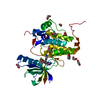 8bioC  8bziC  5nkiS S: Starting model for refinement C: citing same article ( |
|---|---|
| Similar structure data |
-Links
 PDBj
PDBj


- Assembly
| Deposited unit | 
| ||||||||
|---|---|---|---|---|---|---|---|---|---|
| 1 |
| ||||||||
| Unit cell |
|
-Components
| #1: Protein | Mass: 34462.840 Da / Num. of mol.: 1 Source method: isolated from a genetically manipulated source Source: (gene. exp.) Homo sapiens (human) / Gene: EPHA2, ECK / Production host:  Escherichia coli (E. coli) Escherichia coli (E. coli)References: UniProt: P29317, receptor protein-tyrosine kinase | ||||||
|---|---|---|---|---|---|---|---|
| #2: Chemical | ChemComp-EDO / Ethylene glycol  Mass: 62.068 Da / Num. of mol.: 7 / Source method: obtained synthetically / Formula: C2H6O2 Mass: 62.068 Da / Num. of mol.: 7 / Source method: obtained synthetically / Formula: C2H6O2#3: Chemical | ChemComp-QRR / |   Mass: 357.837 Da / Num. of mol.: 1 / Source method: isolated from a natural source / Formula: C18H20ClN5O / Feature type: SUBJECT OF INVESTIGATION Mass: 357.837 Da / Num. of mol.: 1 / Source method: isolated from a natural source / Formula: C18H20ClN5O / Feature type: SUBJECT OF INVESTIGATION#4: Water | ChemComp-HOH / | Water Mass: 18.015 Da / Num. of mol.: 120 / Source method: isolated from a natural source / Formula: H2O Mass: 18.015 Da / Num. of mol.: 120 / Source method: isolated from a natural source / Formula: H2OHas ligand of interest | Y | |
-Experimental details
-Experiment
| Experiment | Method: X-RAY DIFFRACTION / Number of used crystals: 1 |
|---|
- Sample preparation
| Crystal | Density Matthews: 1.97 Å3/Da / Density % sol: 37.59 % |
|---|---|
| Crystal grow | Temperature: 293 K / Method: vapor diffusion, sitting drop / pH: 7.5 Details: 20% PEG3350, 10% ethylene glycol, 0.2M potassium thiocyanate |
-Data collection
| Diffraction | Mean temperature: 100 K / Serial crystal experiment: N |
|---|---|
| Diffraction source | Source: SYNCHROTRON / Site: SLS  / Beamline: X06SA / Wavelength: 0.99999 Å / Beamline: X06SA / Wavelength: 0.99999 Å |
| Detector | Type: DECTRIS EIGER X 16M / Detector: PIXEL / Date: Oct 15, 2022 |
| Radiation | Protocol: SINGLE WAVELENGTH / Monochromatic (M) / Laue (L): M / Scattering type: x-ray |
| Radiation wavelength | Wavelength: 0.99999 Å / Relative weight: 1 |
| Reflection | Resolution: 1.5→38.49 Å / Num. obs: 35835 / % possible obs: 84.2 % / Redundancy: 6.9 % / CC1/2: 0.998 / Net I/σ(I): 14.8 |
| Reflection shell | Resolution: 1.5→1.53 Å / Num. unique obs: 726 / CC1/2: 0.825 |
- Processing
| Software |
| ||||||||||||||||||||||||||||||||||||||||||||||||||||||||||||||||||||||||||||||||||||||||||||||||||||||||||||||||||||||||||||||||||||||||||||||||||||||||||||||||||||||||||||||||||||||
|---|---|---|---|---|---|---|---|---|---|---|---|---|---|---|---|---|---|---|---|---|---|---|---|---|---|---|---|---|---|---|---|---|---|---|---|---|---|---|---|---|---|---|---|---|---|---|---|---|---|---|---|---|---|---|---|---|---|---|---|---|---|---|---|---|---|---|---|---|---|---|---|---|---|---|---|---|---|---|---|---|---|---|---|---|---|---|---|---|---|---|---|---|---|---|---|---|---|---|---|---|---|---|---|---|---|---|---|---|---|---|---|---|---|---|---|---|---|---|---|---|---|---|---|---|---|---|---|---|---|---|---|---|---|---|---|---|---|---|---|---|---|---|---|---|---|---|---|---|---|---|---|---|---|---|---|---|---|---|---|---|---|---|---|---|---|---|---|---|---|---|---|---|---|---|---|---|---|---|---|---|---|---|---|
| Refinement | Method to determine structure: MOLECULAR REPLACEMENT Starting model: 5NKI Resolution: 1.5→38.49 Å / Cor.coef. Fo:Fc: 0.971 / Cor.coef. Fo:Fc free: 0.961 / SU B: 2.449 / SU ML: 0.041 / Cross valid method: THROUGHOUT / ESU R: 0.088 / ESU R Free: 0.072 / Stereochemistry target values: MAXIMUM LIKELIHOOD / Details: HYDROGENS HAVE BEEN ADDED IN THE RIDING POSITIONS
| ||||||||||||||||||||||||||||||||||||||||||||||||||||||||||||||||||||||||||||||||||||||||||||||||||||||||||||||||||||||||||||||||||||||||||||||||||||||||||||||||||||||||||||||||||||||
| Solvent computation | Ion probe radii: 0.8 Å / Shrinkage radii: 0.8 Å / VDW probe radii: 1.2 Å / Solvent model: MASK | ||||||||||||||||||||||||||||||||||||||||||||||||||||||||||||||||||||||||||||||||||||||||||||||||||||||||||||||||||||||||||||||||||||||||||||||||||||||||||||||||||||||||||||||||||||||
| Displacement parameters | Biso mean: 17.938 Å2
| ||||||||||||||||||||||||||||||||||||||||||||||||||||||||||||||||||||||||||||||||||||||||||||||||||||||||||||||||||||||||||||||||||||||||||||||||||||||||||||||||||||||||||||||||||||||
| Refinement step | Cycle: 1 / Resolution: 1.5→38.49 Å
| ||||||||||||||||||||||||||||||||||||||||||||||||||||||||||||||||||||||||||||||||||||||||||||||||||||||||||||||||||||||||||||||||||||||||||||||||||||||||||||||||||||||||||||||||||||||
| Refine LS restraints |
|