 Movie
Movie Controller


+ Open data

- Basic information
| Entry | Database: PDB / ID: 8bia | |||||||||
|---|---|---|---|---|---|---|---|---|---|---|
| Title | Crystal structure of Scribble PDZ1 with PTHR | |||||||||
 Components Components |
| |||||||||
 Keywords Keywords |  PROTEIN BINDING / Scribble / PTHR / PDZ domain / cell polarity PROTEIN BINDING / Scribble / PTHR / PDZ domain / cell polarity | |||||||||
| Function / homology |  Function and homology information Function and homology informationneurotransmitter receptor transport postsynaptic membrane to endosome / extrinsic component of postsynaptic density membrane / establishment of T cell polarity / parathyroid hormone receptor activity / apoptotic process involved in morphogenesis / establishment of apical/basal cell polarity / cochlear nucleus development / synaptic vesicle targeting / Scrib-APC-beta-catenin complex / astrocyte cell migration ...neurotransmitter receptor transport postsynaptic membrane to endosome / extrinsic component of postsynaptic density membrane / establishment of T cell polarity / parathyroid hormone receptor activity / apoptotic process involved in morphogenesis / establishment of apical/basal cell polarity / cochlear nucleus development / synaptic vesicle targeting / Scrib-APC-beta-catenin complex / astrocyte cell migration / polarized epithelial cell differentiation / myelin sheath abaxonal region / neurotransmitter receptor transport, endosome to postsynaptic membrane / protein localization to adherens junction / cell-cell contact zone / mammary gland duct morphogenesis / post-anal tail morphogenesis / G protein-coupled peptide receptor activity / establishment or maintenance of epithelial cell apical/basal polarity / activation of GTPase activity / regulation of postsynaptic neurotransmitter receptor internalization / auditory receptor cell stereocilium organization / Class B/2 (Secretin family receptors) / osteoblast development / RND2 GTPase cycle / RND3 GTPase cycle / positive regulation of inositol phosphate biosynthetic process / positive regulation of receptor recycling / positive chemotaxis / receptor clustering / bone mineralization / RHOJ GTPase cycle / RHOQ GTPase cycle / negative regulation of activated T cell proliferation / G protein-coupled receptor signaling pathway, coupled to cyclic nucleotide second messenger / peptide hormone binding / CDC42 GTPase cycle / synaptic vesicle endocytosis / negative regulation of mitotic cell cycle / immunological synapse / chondrocyte differentiation / signaling adaptor activity / cell maturation / bone resorption / Asymmetric localization of PCP proteins / skeletal system development / neural tube closure / adherens junction / adenylate cyclase-modulating G protein-coupled receptor signaling pathway / wound healing / adenylate cyclase-activating G protein-coupled receptor signaling pathway / cell-cell adhesion / intracellular calcium ion homeostasis / : / cell migration / cell-cell junction / positive regulation of type II interferon production / presynapse / cell junction / lamellipodium / phospholipase C-activating G protein-coupled receptor signaling pathway / G alpha (s) signalling events / basolateral plasma membrane / cell population proliferation / in utero embryonic development / postsynaptic density / cell surface receptor signaling pathway / receptor complex / cadherin binding / positive regulation of apoptotic process / apical plasma membrane / G protein-coupled receptor signaling pathway / negative regulation of cell population proliferation / glutamatergic synapse / positive regulation of cell population proliferation / protein homodimerization activity / extracellular exosome / nucleoplasm / nucleus / plasma membrane / cytoplasmSimilarity search - Function | |||||||||
| Biological species |  Homo sapiens (human) Homo sapiens (human) | |||||||||
| Method | X-RAY DIFFRACTION / SYNCHROTRON / MOLECULAR REPLACEMENT / Resolution: 2.4 Å | |||||||||
 Authors Authors | Stewart, B.Z. / Kvansakul, M. | |||||||||
| Funding support |  Australia, 2items Australia, 2items
| |||||||||
 Citation Citation | Journal: To Be Published Title: Crystal structure of Scribble PDZ1 with human papillomavirus strain 16 E6 peptide Authors: Stewart, B.Z. / Caria, S. / Humbert, P.O. / Kvansakul, M. | |||||||||
| History |
|
- Structure visualization
| Structure viewer | Molecule: MolmilJmol/JSmol |
|---|
- Downloads & links
-Download
| PDBx/mmCIF format | 8bia.cif.gz | 90.7 KB | Display | PDBx/mmCIF format |
|---|---|---|---|---|
| PDB format | pdb8bia.ent.gz | 57.5 KB | Display | PDB format |
| PDBx/mmJSON format | 8bia.json.gz | Tree view | PDBx/mmJSON format | |
| Others |  Other downloads Other downloads |
-Validation report
| Arichive directory | https://data.pdbj.org/pub/pdb/validation_reports/bi/8biaftp://data.pdbj.org/pub/pdb/validation_reports/bi/8bia | HTTPS FTP |
|---|
-Related structure data
| Related structure data |  8b82C 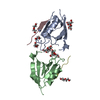 8b87C 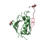 8b8oC 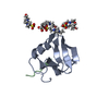 8b9tC 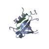 8bj0C 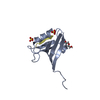 6mtvS S: Starting model for refinement C: citing same article ( |
|---|---|
| Similar structure data |
-Links
 PDBj
PDBj


- Assembly
| Deposited unit | 
| ||||||||||||
|---|---|---|---|---|---|---|---|---|---|---|---|---|---|
| 1 |
| ||||||||||||
| Unit cell |
|
-Components
| #1: Protein | Mass: 12293.823 Da / Num. of mol.: 1 Source method: isolated from a genetically manipulated source Source: (gene. exp.) Homo sapiens (human) / Gene: SCRIB, CRIB1, KIAA0147, LAP4, SCRB1, VARTUL / Production host:  Escherichia coli BL21(DE3) (bacteria) / Strain (production host): Codon+ / References: UniProt: Q14160 Escherichia coli BL21(DE3) (bacteria) / Strain (production host): Codon+ / References: UniProt: Q14160 |
|---|---|
| #2: Protein/peptide | Mass: 1051.126 Da / Num. of mol.: 1 / Source method: obtained synthetically / Source: (synth.) Homo sapiens (human) / References: UniProt: Q03431 |
| #3: Chemical | ChemComp-PO4 / Phosphate  Mass: 94.971 Da / Num. of mol.: 1 / Source method: obtained synthetically / Formula: PO4 Mass: 94.971 Da / Num. of mol.: 1 / Source method: obtained synthetically / Formula: PO4 |
| #4: Water | ChemComp-HOH / Water Mass: 18.015 Da / Num. of mol.: 35 / Source method: isolated from a natural source / Formula: H2O Mass: 18.015 Da / Num. of mol.: 35 / Source method: isolated from a natural source / Formula: H2O |
| Has ligand of interest | N |
-Experimental details
-Experiment
| Experiment | Method: X-RAY DIFFRACTION / Number of used crystals: 1 |
|---|
- Sample preparation
| Crystal | Density Matthews: 3.48 Å3/Da / Density % sol: 64.72 % / Description: triangular diamond prism |
|---|---|
| Crystal grow | Temperature: 293.15 K / Method: vapor diffusion, sitting drop / Details: 0.1 M phosphate/citrate pH 5.2, 43% PEG 300 |
-Data collection
| Diffraction | Mean temperature: 100 K / Serial crystal experiment: N |
|---|---|
| Diffraction source | Source: SYNCHROTRON / Site: Australian Synchrotron / Beamline: MX2 / Wavelength: 0.95372 Å |
| Detector | Type: DECTRIS EIGER X 16M / Detector: PIXEL / Date: Mar 16, 2022 |
| Radiation | Protocol: SINGLE WAVELENGTH / Monochromatic (M) / Laue (L): M / Scattering type: x-ray |
| Radiation wavelength | Wavelength: 0.95372 Å / Relative weight: 1 |
| Reflection | Resolution: 2.4→51.66 Å / Num. obs: 6472 / % possible obs: 99.4 % / Redundancy: 4.4 % / Biso Wilson estimate: 56.57 Å2 / CC1/2: 0.999 / Net I/σ(I): 14.2 |
| Reflection shell | Resolution: 2.4→2.486 Å / Num. unique obs: 623 / CC1/2: 0.891 / % possible all: 98.9 |
- Processing
| Software |
| ||||||||||||||||||||||||||||||||||||||||
|---|---|---|---|---|---|---|---|---|---|---|---|---|---|---|---|---|---|---|---|---|---|---|---|---|---|---|---|---|---|---|---|---|---|---|---|---|---|---|---|---|---|
| Refinement | Method to determine structure: MOLECULAR REPLACEMENT Starting model: 6MTV Resolution: 2.4→51.63 Å / SU ML: 0.1752 / Cross valid method: FREE R-VALUE / σ(F): 1.34 / Phase error: 34.0619 Stereochemistry target values: GeoStd + Monomer Library + CDL v1.2
| ||||||||||||||||||||||||||||||||||||||||
| Solvent computation | Shrinkage radii: 0.9 Å / VDW probe radii: 1.1 Å / Solvent model: FLAT BULK SOLVENT MODEL | ||||||||||||||||||||||||||||||||||||||||
| Displacement parameters | Biso mean: 65.28 Å2 | ||||||||||||||||||||||||||||||||||||||||
| Refinement step | Cycle: LAST / Resolution: 2.4→51.63 Å
| ||||||||||||||||||||||||||||||||||||||||
| Refine LS restraints |
| ||||||||||||||||||||||||||||||||||||||||
| LS refinement shell |
| ||||||||||||||||||||||||||||||||||||||||
| Refinement TLS params. | Method: refined / Origin x: 15.0246235025 Å / Origin y: 27.1692229986 Å / Origin z: -12.4512720899 Å
| ||||||||||||||||||||||||||||||||||||||||
| Refinement TLS group | Selection details: all |
