 Movie
Movie Controller


+ Open data

- Basic information
| Entry | Database: PDB / ID: 8b9h | ||||||
|---|---|---|---|---|---|---|---|
| Title | Crystal structure of JAK2 JH2 in complex with Z902-A3 | ||||||
 Components Components | Tyrosine-protein kinase JAK2 | ||||||
 Keywords Keywords |  TRANSFERASE / Janus kinase / pseudokinase / inhibitor complex / JAK2 / JH2 TRANSFERASE / Janus kinase / pseudokinase / inhibitor complex / JAK2 / JH2 | ||||||
| Function / homology |  Function and homology information Function and homology informationinterleukin-35-mediated signaling pathway / intracellular mineralocorticoid receptor signaling pathway / histone H3Y41 kinase activity / activation of cysteine-type endopeptidase activity involved in apoptotic signaling pathway / positive regulation of growth factor dependent skeletal muscle satellite cell proliferation / symbiont-induced defense-related programmed cell death / mammary gland epithelium development / regulation of postsynapse to nucleus signaling pathway / positive regulation of growth hormone receptor signaling pathway / Signaling by Erythropoietin ...interleukin-35-mediated signaling pathway / intracellular mineralocorticoid receptor signaling pathway / histone H3Y41 kinase activity / activation of cysteine-type endopeptidase activity involved in apoptotic signaling pathway / positive regulation of growth factor dependent skeletal muscle satellite cell proliferation / symbiont-induced defense-related programmed cell death / mammary gland epithelium development / regulation of postsynapse to nucleus signaling pathway / positive regulation of growth hormone receptor signaling pathway / Signaling by Erythropoietin / granulocyte macrophage colony-stimulating factor receptor complex / granulocyte-macrophage colony-stimulating factor signaling pathway / interleukin-12 receptor binding / post-embryonic hemopoiesis / collagen-activated signaling pathway / Erythropoietin activates STAT5 / Erythropoietin activates Phospholipase C gamma (PLCG) / interleukin-5-mediated signaling pathway / response to interleukin-12 / positive regulation of leukocyte proliferation / activation of Janus kinase activity / interleukin-12 receptor complex / interleukin-23 receptor complex / positive regulation of platelet aggregation / tyrosine phosphorylation of STAT protein / Interleukin-23 signaling / positive regulation of MHC class II biosynthetic process / positive regulation of platelet activation / positive regulation of T-helper 17 type immune response / type 1 angiotensin receptor binding / positive regulation of NK T cell proliferation / interleukin-12-mediated signaling pathway / interleukin-3-mediated signaling pathway / regulation of nitric oxide biosynthetic process / acetylcholine receptor binding / cellular response to interleukin-3 / Signaling by Leptin / positive regulation of signaling receptor activity / Interleukin-12 signaling / Interleukin-35 Signalling / Interleukin-27 signaling / IL-6-type cytokine receptor ligand interactions / positive regulation of cell-substrate adhesion / response to hydroperoxide / regulation of receptor signaling pathway via JAK-STAT / growth hormone receptor binding / negative regulation of cardiac muscle cell apoptotic process / positive regulation of epithelial cell apoptotic process / axon regeneration / peptide hormone receptor binding / growth hormone receptor signaling pathway / intrinsic apoptotic signaling pathway in response to oxidative stress / IFNG signaling activates MAPKs / extrinsic component of plasma membrane / Interleukin-20 family signaling / Erythropoietin activates Phosphoinositide-3-kinase (PI3K) / Interleukin-6 signaling / enzyme-linked receptor protein signaling pathway / interleukin-6-mediated signaling pathway / negative regulation of cell-cell adhesion / Prolactin receptor signaling / positive regulation of interleukin-17 production / negative regulation of DNA binding / MAPK3 (ERK1) activation / response to amine / positive regulation of nitric-oxide synthase biosynthetic process / MAPK1 (ERK2) activation / positive regulation of natural killer cell proliferation / mesoderm development / positive regulation of SMAD protein signal transduction / cell surface receptor signaling pathway via JAK-STAT / platelet-derived growth factor receptor signaling pathway / insulin receptor substrate binding / Interleukin-3, Interleukin-5 and GM-CSF signaling / growth hormone receptor signaling pathway via JAK-STAT / response to tumor necrosis factor / Interleukin receptor SHC signaling / phosphatidylinositol 3-kinase binding / type II interferon-mediated signaling pathway / Regulation of IFNG signaling / Erythropoietin activates RAS / Growth hormone receptor signaling / positive regulation of apoptotic signaling pathway / extrinsic apoptotic signaling pathway / Signaling by CSF3 (G-CSF) / positive regulation of tyrosine phosphorylation of STAT protein / positive regulation of vascular associated smooth muscle cell proliferation / positive regulation of T cell proliferation / tumor necrosis factor-mediated signaling pathway / actin filament polymerization / extrinsic component of cytoplasmic side of plasma membrane / SH2 domain binding / cellular response to dexamethasone stimulus / post-translational protein modification / erythrocyte differentiation / Signaling by phosphorylated juxtamembrane, extracellular and kinase domain KIT mutants / positive regulation of interleukin-1 beta production / caveola / endosome lumen / positive regulation of cell differentiationSimilarity search - Function | ||||||
| Biological species |  Homo sapiens (human) Homo sapiens (human) | ||||||
| Method | X-RAY DIFFRACTION / SYNCHROTRON / MOLECULAR REPLACEMENT / Resolution: 1.5 Å | ||||||
 Authors Authors | Haikarainen, T. / Silvennoinen, O. | ||||||
| Funding support |  Finland, 1items Finland, 1items
| ||||||
 Citation Citation | Journal: Pharmaceuticals / Year: 2023 Title: Identification of Novel Small Molecule Ligands for JAK2 Pseudokinase Domain. Authors: Virtanen, A.T. / Haikarainen, T. / Sampathkumar, P. / Palmroth, M. / Liukkonen, S. / Liu, J. / Nekhotiaeva, N. / Hubbard, S.R. / Silvennoinen, O. | ||||||
| History |
|
- Structure visualization
| Structure viewer | Molecule: MolmilJmol/JSmol |
|---|
- Downloads & links
-Download
| PDBx/mmCIF format | 8b9h.cif.gz | 201.8 KB | Display | PDBx/mmCIF format |
|---|---|---|---|---|
| PDB format | pdb8b9h.ent.gz | 140.1 KB | Display | PDB format |
| PDBx/mmJSON format | 8b9h.json.gz | Tree view | PDBx/mmJSON format | |
| Others |  Other downloads Other downloads |
-Validation report
| Arichive directory | https://data.pdbj.org/pub/pdb/validation_reports/b9/8b9hftp://data.pdbj.org/pub/pdb/validation_reports/b9/8b9h | HTTPS FTP |
|---|
-Related structure data
| Related structure data |  8b8nC 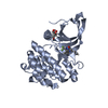 8b8uC  8b99C 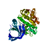 8b9eC 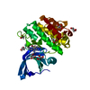 8ba2C 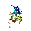 8ba3C 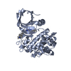 8ba4C 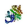 8babC  8bakC  8ex0C  8ex1C 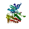 8ex2C  4fvrS S: Starting model for refinement C: citing same article ( |
|---|---|
| Similar structure data |
-Links
 PDBj
PDBj


- Assembly
| Deposited unit | 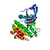
| ||||||||||||
|---|---|---|---|---|---|---|---|---|---|---|---|---|---|
| 1 |
| ||||||||||||
| Unit cell |
|
-Components
| #1: Protein | Mass: 33120.961 Da / Num. of mol.: 1 / Mutation: W659A, W777A, F794H Source method: isolated from a genetically manipulated source Source: (gene. exp.) Homo sapiens (human) / Gene: JAK2 / Production host:   Spodoptera frugiperda (fall armyworm) Spodoptera frugiperda (fall armyworm)References: UniProt: O60674, non-specific protein-tyrosine kinase | ||||
|---|---|---|---|---|---|
| #2: Chemical | ChemComp-Q7F / 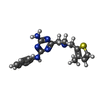  Mass: 340.446 Da / Num. of mol.: 1 / Source method: obtained synthetically / Formula: C17H20N6S / Feature type: SUBJECT OF INVESTIGATION Mass: 340.446 Da / Num. of mol.: 1 / Source method: obtained synthetically / Formula: C17H20N6S / Feature type: SUBJECT OF INVESTIGATION | ||||
| #3: Chemical | ChemComp-GOL / Glycerol  Mass: 92.094 Da / Num. of mol.: 4 / Source method: obtained synthetically / Formula: C3H8O3 Mass: 92.094 Da / Num. of mol.: 4 / Source method: obtained synthetically / Formula: C3H8O3#4: Water | ChemComp-HOH / | Water Mass: 18.015 Da / Num. of mol.: 175 / Source method: isolated from a natural source / Formula: H2O Mass: 18.015 Da / Num. of mol.: 175 / Source method: isolated from a natural source / Formula: H2OHas ligand of interest | Y | |
-Experimental details
-Experiment
| Experiment | Method: X-RAY DIFFRACTION / Number of used crystals: 1 |
|---|
- Sample preparation
| Crystal | Density Matthews: 2.18 Å3/Da / Density % sol: 43.63 % |
|---|---|
| Crystal grow | Temperature: 277 K / Method: vapor diffusion, hanging drop / pH: 8 / Details: 0.1M Tris pH 8, 20% PEG4000, 0.2M Na-acetate |
-Data collection
| Diffraction | Mean temperature: 100 K / Serial crystal experiment: N |
|---|---|
| Diffraction source | Source: SYNCHROTRON / Site: Diamond  / Beamline: I03 / Wavelength: 0.97625 Å / Beamline: I03 / Wavelength: 0.97625 Å |
| Detector | Type: DECTRIS EIGER2 XE 16M / Detector: PIXEL / Date: Nov 24, 2020 |
| Radiation | Protocol: SINGLE WAVELENGTH / Monochromatic (M) / Laue (L): M / Scattering type: x-ray |
| Radiation wavelength | Wavelength: 0.97625 Å / Relative weight: 1 |
| Reflection | Resolution: 1.5→57.319 Å / Num. obs: 86234 / % possible obs: 95.9 % / Redundancy: 3.6 % / Biso Wilson estimate: 22.55 Å2 / CC1/2: 0.999 / Net I/σ(I): 12.23 |
| Reflection shell | Resolution: 1.5→1.59 Å / Redundancy: 3.3 % / Mean I/σ(I) obs: 1.05 / Num. unique obs: 12884 / CC1/2: 0.68 / % possible all: 88.7 |
- Processing
| Software |
| |||||||||||||||||||||||||||||||||||||||||||||||||||||||||||||||||||||||||||||||||||||||||||||||||||||||||||||||||||||||||||||||||||||||||||||||||||||||||||||||||||||||||||||||||||||||||||||||||||||||||||||||||||||||||
|---|---|---|---|---|---|---|---|---|---|---|---|---|---|---|---|---|---|---|---|---|---|---|---|---|---|---|---|---|---|---|---|---|---|---|---|---|---|---|---|---|---|---|---|---|---|---|---|---|---|---|---|---|---|---|---|---|---|---|---|---|---|---|---|---|---|---|---|---|---|---|---|---|---|---|---|---|---|---|---|---|---|---|---|---|---|---|---|---|---|---|---|---|---|---|---|---|---|---|---|---|---|---|---|---|---|---|---|---|---|---|---|---|---|---|---|---|---|---|---|---|---|---|---|---|---|---|---|---|---|---|---|---|---|---|---|---|---|---|---|---|---|---|---|---|---|---|---|---|---|---|---|---|---|---|---|---|---|---|---|---|---|---|---|---|---|---|---|---|---|---|---|---|---|---|---|---|---|---|---|---|---|---|---|---|---|---|---|---|---|---|---|---|---|---|---|---|---|---|---|---|---|---|---|---|---|---|---|---|---|---|---|---|---|---|---|---|---|---|
| Refinement | Method to determine structure: MOLECULAR REPLACEMENT Starting model: 4fvr Resolution: 1.5→41.26 Å / SU ML: 0.2056 / Cross valid method: FREE R-VALUE / σ(F): 1.36 / Phase error: 32.5386 Stereochemistry target values: GeoStd + Monomer Library + CDL v1.2
| |||||||||||||||||||||||||||||||||||||||||||||||||||||||||||||||||||||||||||||||||||||||||||||||||||||||||||||||||||||||||||||||||||||||||||||||||||||||||||||||||||||||||||||||||||||||||||||||||||||||||||||||||||||||||
| Solvent computation | Shrinkage radii: 0.9 Å / VDW probe radii: 1.11 Å / Solvent model: FLAT BULK SOLVENT MODEL | |||||||||||||||||||||||||||||||||||||||||||||||||||||||||||||||||||||||||||||||||||||||||||||||||||||||||||||||||||||||||||||||||||||||||||||||||||||||||||||||||||||||||||||||||||||||||||||||||||||||||||||||||||||||||
| Displacement parameters | Biso mean: 37.88 Å2 | |||||||||||||||||||||||||||||||||||||||||||||||||||||||||||||||||||||||||||||||||||||||||||||||||||||||||||||||||||||||||||||||||||||||||||||||||||||||||||||||||||||||||||||||||||||||||||||||||||||||||||||||||||||||||
| Refinement step | Cycle: LAST / Resolution: 1.5→41.26 Å
| |||||||||||||||||||||||||||||||||||||||||||||||||||||||||||||||||||||||||||||||||||||||||||||||||||||||||||||||||||||||||||||||||||||||||||||||||||||||||||||||||||||||||||||||||||||||||||||||||||||||||||||||||||||||||
| Refine LS restraints |
| |||||||||||||||||||||||||||||||||||||||||||||||||||||||||||||||||||||||||||||||||||||||||||||||||||||||||||||||||||||||||||||||||||||||||||||||||||||||||||||||||||||||||||||||||||||||||||||||||||||||||||||||||||||||||
| LS refinement shell |
|
