 Movie
Movie Controller

[English] 日本語
 Yorodumi
- PDB-8b7x: X-ray structure of the CeuE Homologue from Geobacillus stearother... -
+ Open data

- Basic information
| Entry | Database: PDB / ID: 8b7x | ||||||
|---|---|---|---|---|---|---|---|
| Title | X-ray structure of the CeuE Homologue from Geobacillus stearothermophilus - apo form. | ||||||
 Components Components | Siderophore ABC transporter substrate-binding protein | ||||||
 Keywords Keywords | METAL TRANSPORT /  Periplasmic / Siderophore binding / Bacterial / apo protein. Periplasmic / Siderophore binding / Bacterial / apo protein. | ||||||
| Function / homology | FatB domain / ABC transporter periplasmic binding domain / Periplasmic binding protein / Iron siderophore/cobalamin periplasmic-binding domain profile. / Prokaryotic membrane lipoprotein lipid attachment site profile. / iron ion transport / Chem-JEF / Siderophore ABC transporter substrate-binding protein Function and homology information Function and homology information | ||||||
| Biological species |   Geobacillus stearothermophilus (bacteria) Geobacillus stearothermophilus (bacteria) | ||||||
| Method | X-RAY DIFFRACTION / SYNCHROTRON / MOLECULAR REPLACEMENT / Resolution: 1.42 Å | ||||||
 Authors Authors | Wilson, K.S. / Duhme-Klair, A.K. / Blagova, E.V. / Bennett, M. | ||||||
| Funding support |  United Kingdom, 1items United Kingdom, 1items
| ||||||
 Citation Citation | Journal: Acta Crystallogr D Struct Biol / Year: 2023 Title: Thermostable homologues of the periplasmic siderophore-binding protein CeuE from Geobacillus stearothermophilus and Parageobacillus thermoglucosidasius. Authors: Blagova, E.V. / Miller, A.H. / Bennett, M. / Booth, R.L. / Dodson, E.J. / Duhme-Klair, A.K. / Wilson, K.S. | ||||||
| History |
|
- Structure visualization
| Structure viewer | Molecule: MolmilJmol/JSmol |
|---|
- Downloads & links
-Download
| PDBx/mmCIF format | 8b7x.cif.gz | 299.1 KB | Display | PDBx/mmCIF format |
|---|---|---|---|---|
| PDB format | pdb8b7x.ent.gz | 185.9 KB | Display | PDB format |
| PDBx/mmJSON format | 8b7x.json.gz | Tree view | PDBx/mmJSON format | |
| Others |  Other downloads Other downloads |
-Validation report
| Arichive directory | https://data.pdbj.org/pub/pdb/validation_reports/b7/8b7xftp://data.pdbj.org/pub/pdb/validation_reports/b7/8b7x | HTTPS FTP |
|---|
-Related structure data
| Related structure data |  8bawC  8baxC  8bf6C  8bj9C  8bnwC  3zkwS S: Starting model for refinement C: citing same article ( |
|---|---|
| Similar structure data |
-Links
 PDBj
PDBj


- Assembly
| Deposited unit | 
| ||||||||
|---|---|---|---|---|---|---|---|---|---|
| 1 |
| ||||||||
| Unit cell |
|
-Components
| #1: Protein | Mass: 33385.930 Da / Num. of mol.: 1 Source method: isolated from a genetically manipulated source Source: (gene. exp.) Geobacillus stearothermophilus (bacteria)Gene: EPB69_07310 / Production host: Escherichia coli BL21(DE3) (bacteria) / References: UniProt: A0A857MR34 |
|---|---|
| #2: Chemical | ChemComp-JEF / 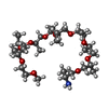  Mass: 597.822 Da / Num. of mol.: 1 / Source method: obtained synthetically / Formula: C30H63NO10 Mass: 597.822 Da / Num. of mol.: 1 / Source method: obtained synthetically / Formula: C30H63NO10 |
| #3: Chemical | ChemComp-SO4 / Sulfate  Mass: 96.063 Da / Num. of mol.: 1 / Source method: isolated from a natural source / Formula: SO4 Mass: 96.063 Da / Num. of mol.: 1 / Source method: isolated from a natural source / Formula: SO4 |
| #4: Water | ChemComp-HOH / Water Mass: 18.015 Da / Num. of mol.: 56 / Source method: isolated from a natural source / Formula: H2O Mass: 18.015 Da / Num. of mol.: 56 / Source method: isolated from a natural source / Formula: H2O |
| Has ligand of interest | N |
-Experimental details
-Experiment
| Experiment | Method: X-RAY DIFFRACTION / Number of used crystals: 1 |
|---|
- Sample preparation
| Crystal | Density % sol: 26.11 % |
|---|---|
| Crystal grow | Temperature: 293 K / Method: vapor diffusion, hanging drop Details: Index D3: 0.1M HEPES, pH 7.0, 30% Jeffamine ED 2001, pH 7.0. No cryo. |
-Data collection
| Diffraction | Mean temperature: 100 K / Serial crystal experiment: N | |||||||||||||||||||||
|---|---|---|---|---|---|---|---|---|---|---|---|---|---|---|---|---|---|---|---|---|---|---|
| Diffraction source | Source: SYNCHROTRON / Site: Diamond / Beamline: I03 / Wavelength: 0.97622 Å | |||||||||||||||||||||
| Detector | Type: DECTRIS PILATUS3 6M / Detector: PIXEL / Date: Nov 25, 2018 | |||||||||||||||||||||
| Radiation | Protocol: SINGLE WAVELENGTH / Monochromatic (M) / Laue (L): M / Scattering type: x-ray | |||||||||||||||||||||
| Radiation wavelength | Wavelength: 0.97622 Å / Relative weight: 1 | |||||||||||||||||||||
| Reflection | Resolution: 1.42→32.225 Å / Num. obs: 37722 / % possible obs: 94.9 % / Redundancy: 2 % / CC1/2: 0.998 / Rmerge(I) obs: 0.033 / Rpim(I) all: 0.033 / Rrim(I) all: 0.046 / Net I/σ(I): 5.8 | |||||||||||||||||||||
| Reflection shell | Diffraction-ID: 1
|
- Processing
| Software |
| |||||||||||||||||||||||||||||||||||||||||||||||||||||||||||||||||||||||||||||||||||||||||||||||||||||||||||||||||||||||||||||||||||||||||||||||||||||||||||||||||||||||||||||||||||||||||||||||||||||||||||||||||||||||||||||||||||||||
|---|---|---|---|---|---|---|---|---|---|---|---|---|---|---|---|---|---|---|---|---|---|---|---|---|---|---|---|---|---|---|---|---|---|---|---|---|---|---|---|---|---|---|---|---|---|---|---|---|---|---|---|---|---|---|---|---|---|---|---|---|---|---|---|---|---|---|---|---|---|---|---|---|---|---|---|---|---|---|---|---|---|---|---|---|---|---|---|---|---|---|---|---|---|---|---|---|---|---|---|---|---|---|---|---|---|---|---|---|---|---|---|---|---|---|---|---|---|---|---|---|---|---|---|---|---|---|---|---|---|---|---|---|---|---|---|---|---|---|---|---|---|---|---|---|---|---|---|---|---|---|---|---|---|---|---|---|---|---|---|---|---|---|---|---|---|---|---|---|---|---|---|---|---|---|---|---|---|---|---|---|---|---|---|---|---|---|---|---|---|---|---|---|---|---|---|---|---|---|---|---|---|---|---|---|---|---|---|---|---|---|---|---|---|---|---|---|---|---|---|---|---|---|---|---|---|---|---|---|---|---|---|---|
| Refinement | Method to determine structure: MOLECULAR REPLACEMENT Starting model: 3zkw Resolution: 1.42→32.225 Å / Cor.coef. Fo:Fc: 0.978 / Cor.coef. Fo:Fc free: 0.959 / WRfactor Rfree: 0.249 / WRfactor Rwork: 0.179 / SU B: 6.73 / SU ML: 0.105 / Average fsc free: 0.9556 / Average fsc work: 0.9828 / Cross valid method: FREE R-VALUE / ESU R: 0.094 / ESU R Free: 0.085 Details: Hydrogens have been added in their riding positions
| |||||||||||||||||||||||||||||||||||||||||||||||||||||||||||||||||||||||||||||||||||||||||||||||||||||||||||||||||||||||||||||||||||||||||||||||||||||||||||||||||||||||||||||||||||||||||||||||||||||||||||||||||||||||||||||||||||||||
| Solvent computation | Ion probe radii: 0.8 Å / Shrinkage radii: 0.8 Å / VDW probe radii: 1.2 Å / Solvent model: MASK BULK SOLVENT | |||||||||||||||||||||||||||||||||||||||||||||||||||||||||||||||||||||||||||||||||||||||||||||||||||||||||||||||||||||||||||||||||||||||||||||||||||||||||||||||||||||||||||||||||||||||||||||||||||||||||||||||||||||||||||||||||||||||
| Displacement parameters | Biso mean: 28.729 Å2
| |||||||||||||||||||||||||||||||||||||||||||||||||||||||||||||||||||||||||||||||||||||||||||||||||||||||||||||||||||||||||||||||||||||||||||||||||||||||||||||||||||||||||||||||||||||||||||||||||||||||||||||||||||||||||||||||||||||||
| Refinement step | Cycle: LAST / Resolution: 1.42→32.225 Å
| |||||||||||||||||||||||||||||||||||||||||||||||||||||||||||||||||||||||||||||||||||||||||||||||||||||||||||||||||||||||||||||||||||||||||||||||||||||||||||||||||||||||||||||||||||||||||||||||||||||||||||||||||||||||||||||||||||||||
| Refine LS restraints |
| |||||||||||||||||||||||||||||||||||||||||||||||||||||||||||||||||||||||||||||||||||||||||||||||||||||||||||||||||||||||||||||||||||||||||||||||||||||||||||||||||||||||||||||||||||||||||||||||||||||||||||||||||||||||||||||||||||||||
| LS refinement shell | Refine-ID: X-RAY DIFFRACTION / Total num. of bins used: 20
|
