 Movie
Movie Controller

[English] 日本語
 Yorodumi
- PDB-8b3p: CryoEM structure of the round tip (proteins pVII/pVIII/pIX) from ... -
+ Open data

- Basic information
| Entry | Database: PDB / ID: 8b3p | ||||||
|---|---|---|---|---|---|---|---|
| Title | CryoEM structure of the round tip (proteins pVII/pVIII/pIX) from the f1 filamentous bacteriophage | ||||||
 Components Components |
| ||||||
 Keywords Keywords |  VIRUS / Viral proteins / assembly VIRUS / Viral proteins / assembly | ||||||
| Function / homology |  Function and homology information Function and homology information | ||||||
| Biological species |  Enterobacteria phage f1 (virus) Enterobacteria phage f1 (virus) | ||||||
| Method | ELECTRON MICROSCOPY / single particle reconstruction / cryo EM / Resolution: 2.81 Å | ||||||
 Authors Authors | Conners, R. / McLaren, M. / Gold, V.A.M. | ||||||
| Funding support |  United Kingdom, 1items United Kingdom, 1items
| ||||||
 Citation Citation | Journal: Nat Commun / Year: 2023 Title: Cryo-electron microscopy of the f1 filamentous phage reveals insights into viral infection and assembly. Authors: Rebecca Conners / Rayén Ignacia León-Quezada / Mathew McLaren / Nicholas J Bennett / Bertram Daum / Jasna Rakonjac / Vicki A M Gold /  Abstract: Phages are viruses that infect bacteria and dominate every ecosystem on our planet. As well as impacting microbial ecology, physiology and evolution, phages are exploited as tools in molecular ...Phages are viruses that infect bacteria and dominate every ecosystem on our planet. As well as impacting microbial ecology, physiology and evolution, phages are exploited as tools in molecular biology and biotechnology. This is particularly true for the Ff (f1, fd or M13) phages, which represent a widely distributed group of filamentous viruses. Over nearly five decades, Ffs have seen an extraordinary range of applications, yet the complete structure of the phage capsid and consequently the mechanisms of infection and assembly remain largely mysterious. In this work, we use cryo-electron microscopy and a highly efficient system for production of short Ff-derived nanorods to determine a structure of a filamentous virus including the tips. We show that structure combined with mutagenesis can identify phage domains that are important in bacterial attack and for release of new progeny, allowing new models to be proposed for the phage lifecycle. | ||||||
| History |
|
- Structure visualization
| Structure viewer | Molecule: MolmilJmol/JSmol |
|---|
- Downloads & links
-Download
| PDBx/mmCIF format | 8b3p.cif.gz | 430.3 KB | Display | PDBx/mmCIF format |
|---|---|---|---|---|
| PDB format | pdb8b3p.ent.gz | Display | PDB format | |
| PDBx/mmJSON format | 8b3p.json.gz | Tree view | PDBx/mmJSON format | |
| Others |  Other downloads Other downloads |
-Validation report
| Arichive directory | https://data.pdbj.org/pub/pdb/validation_reports/b3/8b3pftp://data.pdbj.org/pub/pdb/validation_reports/b3/8b3p | HTTPS FTP |
|---|
-Related structure data
| Related structure data | 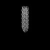 15832MC 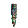 8b3oC 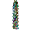 8b3qC M: map data used to model this data C: citing same article ( |
|---|---|
| Similar structure data |


-Links
 PDBj
PDBj- Assembly
| Deposited unit | 
|
|---|---|
| 1 |
|
-Components
| #1: Protein/peptide | Mass: 3603.215 Da / Num. of mol.: 5 Source method: isolated from a genetically manipulated source Source: (gene. exp.) Enterobacteria phage f1 (virus) / Gene: VII / Production host:  Escherichia coli (E. coli) / References: UniProt: P69534 Escherichia coli (E. coli) / References: UniProt: P69534#2: Protein/peptide | Mass: 3655.270 Da / Num. of mol.: 5 Source method: isolated from a genetically manipulated source Source: (gene. exp.) Enterobacteria phage f1 (virus) / Gene: IX / Production host: Escherichia coli (E. coli) / References: UniProt: P69537#3: Protein/peptide | / Coat protein B / Gene 8 protein / G8P / Major coat proteinMass: 5212.021 Da / Num. of mol.: 45 / Mutation: Y44M Source method: isolated from a genetically manipulated source Source: (gene. exp.) Enterobacteria phage f1 (virus) / Gene: VIII / Production host: Escherichia coli (E. coli) / References: UniProt: P69540 |
|---|
-Experimental details
-Experiment
| Experiment | Method: ELECTRON MICROSCOPY |
|---|---|
| EM experiment | Aggregation state: PARTICLE / 3D reconstruction method: single particle reconstruction |
- Sample preparation
| Component | Name: Enterobacteria phage f1 / Type: VIRUS / Entity ID: all / Source: RECOMBINANT |
|---|---|
| Source (natural) | Organism: Enterobacteria phage f1 (virus) |
| Source (recombinant) | Organism: Escherichia coli (E. coli) |
| Details of virus | Empty: NO / Enveloped: NO / Isolate: OTHER / Type: VIRION |
| Natural host | Organism: Escherichia coli / Strain: F+ strains |
| Buffer solution | pH: 8 |
| Specimen | Embedding applied: NO / Shadowing applied: NO / Staining applied: NO / Vitrification applied: YES |
| Specimen support | Grid material: COPPER / Grid mesh size: 300 divisions/in. / Grid type: Quantifoil R1.2/1.3 |
| Vitrification | Instrument: FEI VITROBOT MARK IV / Cryogen name: ETHANE / Humidity: 100 % / Chamber temperature: 277 K Details: Wait time 5 sec., drain time 0 sec., blot force 0, blot time 4 sec. |
- Electron microscopy imaging
| Experimental equipment |  Model: Titan Krios / Image courtesy: FEI Company |
|---|---|
| Microscopy | Model: TFS KRIOS |
| Electron gun | Electron source: FIELD EMISSION GUN / Accelerating voltage: 300 kV / Illumination mode: FLOOD BEAM |
| Electron lens | Mode: BRIGHT FIELDBright-field microscopy / Nominal defocus max: 2500 nm / Nominal defocus min: 1300 nm |
| Image recording | Electron dose: 40 e/Å2 / Film or detector model: GATAN K3 BIOQUANTUM (6k x 4k) |
- Processing
| Software | Name: REFMAC / Version: 5.8.0253 / Classification: refinement / Contact author: Garib N. Murshudov / Contact author email: garib[at]mrc-lmb.cam.ac.uk / Date: Jun 20, 2019 Description: (un)restrained refinement or idealisation of macromolecular structures | |||||||||||||||||||||||||||||||||||||||||||||||||||||||||||||||||||||||||||||||||||||||||||||||||||||||||||||||||||||||||||||||||||||||||||||||||||
|---|---|---|---|---|---|---|---|---|---|---|---|---|---|---|---|---|---|---|---|---|---|---|---|---|---|---|---|---|---|---|---|---|---|---|---|---|---|---|---|---|---|---|---|---|---|---|---|---|---|---|---|---|---|---|---|---|---|---|---|---|---|---|---|---|---|---|---|---|---|---|---|---|---|---|---|---|---|---|---|---|---|---|---|---|---|---|---|---|---|---|---|---|---|---|---|---|---|---|---|---|---|---|---|---|---|---|---|---|---|---|---|---|---|---|---|---|---|---|---|---|---|---|---|---|---|---|---|---|---|---|---|---|---|---|---|---|---|---|---|---|---|---|---|---|---|---|---|---|
| EM software |
| |||||||||||||||||||||||||||||||||||||||||||||||||||||||||||||||||||||||||||||||||||||||||||||||||||||||||||||||||||||||||||||||||||||||||||||||||||
| CTF correction | Type: PHASE FLIPPING AND AMPLITUDE CORRECTION | |||||||||||||||||||||||||||||||||||||||||||||||||||||||||||||||||||||||||||||||||||||||||||||||||||||||||||||||||||||||||||||||||||||||||||||||||||
| Symmetry | Point symmetry: C5 (5 fold cyclic) | |||||||||||||||||||||||||||||||||||||||||||||||||||||||||||||||||||||||||||||||||||||||||||||||||||||||||||||||||||||||||||||||||||||||||||||||||||
| 3D reconstruction | Resolution: 2.81 Å / Resolution method: FSC 0.143 CUT-OFF / Num. of particles: 255372 / Symmetry type: POINT | |||||||||||||||||||||||||||||||||||||||||||||||||||||||||||||||||||||||||||||||||||||||||||||||||||||||||||||||||||||||||||||||||||||||||||||||||||
| Atomic model building | Protocol: OTHER | |||||||||||||||||||||||||||||||||||||||||||||||||||||||||||||||||||||||||||||||||||||||||||||||||||||||||||||||||||||||||||||||||||||||||||||||||||
| Refinement | Resolution: 2.81→222.442 Å / Cor.coef. Fo:Fc: 0.957 / WRfactor Rwork: 0.289 / SU B: 12.907 / SU ML: 0.226 / Average fsc overall: 0.7375 / Average fsc work: 0.7375 / ESU R: 0.447 Details: Hydrogens have been added in their riding positions
| |||||||||||||||||||||||||||||||||||||||||||||||||||||||||||||||||||||||||||||||||||||||||||||||||||||||||||||||||||||||||||||||||||||||||||||||||||
| Solvent computation | Solvent model: BABINET MODEL | |||||||||||||||||||||||||||||||||||||||||||||||||||||||||||||||||||||||||||||||||||||||||||||||||||||||||||||||||||||||||||||||||||||||||||||||||||
| Displacement parameters | Biso mean: 104.571 Å2
| |||||||||||||||||||||||||||||||||||||||||||||||||||||||||||||||||||||||||||||||||||||||||||||||||||||||||||||||||||||||||||||||||||||||||||||||||||
| Refine LS restraints |
| |||||||||||||||||||||||||||||||||||||||||||||||||||||||||||||||||||||||||||||||||||||||||||||||||||||||||||||||||||||||||||||||||||||||||||||||||||
| LS refinement shell | Refine-ID: ELECTRON MICROSCOPY / Num. reflection Rfree: 0 / Total num. of bins used: 20 / % reflection obs: 100 %
|
