 Movie
Movie Controller

[English] 日本語
 Yorodumi
- PDB-8awz: Crystal structure of Trametes versicolor glutathione transferase ... -
+ Open data

- Basic information
| Entry | Database: PDB / ID: 8awz | ||||||
|---|---|---|---|---|---|---|---|
| Title | Crystal structure of Trametes versicolor glutathione transferase Omega 3S in complex with dinitrosyl glutathionyl iron complex (DNGIC) | ||||||
 Components Components | Glutathione transferase Glutathione S-transferase Glutathione S-transferase | ||||||
 Keywords Keywords | TRANSFERASE / glutathione transferase / GSTO / detoxification | ||||||
| Function / homology |  Function and homology information Function and homology information | ||||||
| Biological species |  Trametes versicolor (fungus) Trametes versicolor (fungus) | ||||||
| Method | X-RAY DIFFRACTION / SYNCHROTRON / MOLECULAR REPLACEMENT / Resolution: 1.549 Å | ||||||
 Authors Authors | Schwartz, M. / Didierjean, C. | ||||||
| Funding support |  France, 1items France, 1items
| ||||||
 Citation Citation | Journal: Biochem.Biophys.Res.Commun. / Year: 2023 Title: Structural insights into the interactions of glutathione transferases with a nitric oxide carrier and sodium nitroprusside. Authors: Schwartz, M. / Perrot, T. / Beurton, J. / Zannini, F. / Morel-Rouhier, M. / Gelhaye, E. / Neiers, F. / Schaniel, D. / Favier, F. / Jacquot, J.P. / Leroy, P. / Clarot, I. / Boudier, A. / Didierjean, C. | ||||||
| History |
|
- Structure visualization
| Structure viewer | Molecule: MolmilJmol/JSmol |
|---|
- Downloads & links
-Download
| PDBx/mmCIF format | 8awz.cif.gz | 126.6 KB | Display | PDBx/mmCIF format |
|---|---|---|---|---|
| PDB format | pdb8awz.ent.gz | 95.9 KB | Display | PDB format |
| PDBx/mmJSON format | 8awz.json.gz | Tree view | PDBx/mmJSON format | |
| Others |  Other downloads Other downloads |
-Validation report
| Arichive directory | https://data.pdbj.org/pub/pdb/validation_reports/aw/8awzftp://data.pdbj.org/pub/pdb/validation_reports/aw/8awz | HTTPS FTP |
|---|
-Related structure data
| Related structure data |  8ax0C 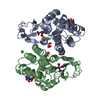 8ax1C  8ax2C  6f43S S: Starting model for refinement C: citing same article ( |
|---|---|
| Similar structure data |
-Links
 PDBj
PDBj


- Assembly
| Deposited unit | 
| ||||||||
|---|---|---|---|---|---|---|---|---|---|
| 1 |
| ||||||||
| Unit cell |
|
-Components
-Protein , 1 types, 2 molecules AB
| #1: Protein | Glutathione S-transferase / Glutathione transferase Omega 3S Mass: 27526.504 Da / Num. of mol.: 2 Source method: isolated from a genetically manipulated source Source: (gene. exp.) Trametes versicolor (fungus) / Production host:  Escherichia coli (E. coli) / References: UniProt: A0A384E145, glutathione transferase Escherichia coli (E. coli) / References: UniProt: A0A384E145, glutathione transferase |
|---|
-Non-polymers , 8 types, 605 molecules 


| #2: Chemical | ChemComp-PG4 / Polyethylene glycol Mass: 194.226 Da / Num. of mol.: 1 / Source method: obtained synthetically / Formula: C8H18O5 / Comment: precipitant*YM Mass: 194.226 Da / Num. of mol.: 1 / Source method: obtained synthetically / Formula: C8H18O5 / Comment: precipitant*YM | ||||||||||||
|---|---|---|---|---|---|---|---|---|---|---|---|---|---|
| #3: Chemical | Glycerol Mass: 92.094 Da / Num. of mol.: 2 / Source method: obtained synthetically / Formula: C3H8O3 Mass: 92.094 Da / Num. of mol.: 2 / Source method: obtained synthetically / Formula: C3H8O3#4: Chemical | Iron Mass: 55.845 Da / Num. of mol.: 2 / Source method: obtained synthetically / Formula: Fe / Feature type: SUBJECT OF INVESTIGATION Mass: 55.845 Da / Num. of mol.: 2 / Source method: obtained synthetically / Formula: Fe / Feature type: SUBJECT OF INVESTIGATION#5: Chemical | ChemComp-NO / Nitric oxide Mass: 30.006 Da / Num. of mol.: 4 / Source method: obtained synthetically / Formula: NO / Feature type: SUBJECT OF INVESTIGATION Mass: 30.006 Da / Num. of mol.: 4 / Source method: obtained synthetically / Formula: NO / Feature type: SUBJECT OF INVESTIGATION#6: Chemical | Glutathione Mass: 307.323 Da / Num. of mol.: 2 / Source method: obtained synthetically / Formula: C10H17N3O6S / Feature type: SUBJECT OF INVESTIGATION Mass: 307.323 Da / Num. of mol.: 2 / Source method: obtained synthetically / Formula: C10H17N3O6S / Feature type: SUBJECT OF INVESTIGATION#7: Chemical | ChemComp-CA / |  Mass: 40.078 Da / Num. of mol.: 1 / Source method: obtained synthetically / Formula: Ca Mass: 40.078 Da / Num. of mol.: 1 / Source method: obtained synthetically / Formula: Ca#8: Chemical | ChemComp-PEG / | Diethylene glycol Mass: 106.120 Da / Num. of mol.: 1 / Source method: obtained synthetically / Formula: C4H10O3 Mass: 106.120 Da / Num. of mol.: 1 / Source method: obtained synthetically / Formula: C4H10O3#9: Water | ChemComp-HOH / | WaterMass: 18.015 Da / Num. of mol.: 592 / Source method: isolated from a natural source / Formula: H2O |
-Details
| Has ligand of interest | Y |
|---|
-Experimental details
-Experiment
| Experiment | Method: X-RAY DIFFRACTION / Number of used crystals: 1 |
|---|
- Sample preparation
| Crystal | Density Matthews: 2.56 Å3/Da / Density % sol: 51.89 % |
|---|---|
| Crystal grow | Temperature: 277 K / Method: microbatch Details: 30 % PEG 400, 0.2 M Calcium Acetate, 0.1 M Acetate Buffer, pH 4.5 |
-Data collection
| Diffraction | Mean temperature: 100 K / Serial crystal experiment: N |
|---|---|
| Diffraction source | Source: SYNCHROTRON / Site: ESRF / Beamline: BM30A / Wavelength: 0.981401 Å |
| Detector | Type: ADSC QUANTUM 315r / Detector: CCD / Date: Apr 4, 2017 |
| Radiation | Protocol: SINGLE WAVELENGTH / Monochromatic (M) / Laue (L): M / Scattering type: x-ray |
| Radiation wavelength | Wavelength: 0.981401 Å / Relative weight: 1 |
| Reflection | Resolution: 1.549→45.36 Å / Num. obs: 82204 / % possible obs: 99.3 % / Redundancy: 4.4 % / Biso Wilson estimate: 15.68 Å2 / CC1/2: 1 / Rmerge(I) obs: 0.065 / Rrim(I) all: 0.074 / Net I/σ(I): 15.04 |
| Reflection shell | Resolution: 1.549→1.59 Å / Rmerge(I) obs: 0.744 / Mean I/σ(I) obs: 1.62 / Num. unique obs: 5668 / CC1/2: 0.58 / Rrim(I) all: 0.873 |
- Processing
| Software |
| ||||||||||||||||||||||||||||||||||||||||||||||||||||||||||||||||||||||||||||||||||||||||||||||||||||||||||||||||||||||||||||||||||||||||||||||||||||||||||||||||||||||||||||||||||||
|---|---|---|---|---|---|---|---|---|---|---|---|---|---|---|---|---|---|---|---|---|---|---|---|---|---|---|---|---|---|---|---|---|---|---|---|---|---|---|---|---|---|---|---|---|---|---|---|---|---|---|---|---|---|---|---|---|---|---|---|---|---|---|---|---|---|---|---|---|---|---|---|---|---|---|---|---|---|---|---|---|---|---|---|---|---|---|---|---|---|---|---|---|---|---|---|---|---|---|---|---|---|---|---|---|---|---|---|---|---|---|---|---|---|---|---|---|---|---|---|---|---|---|---|---|---|---|---|---|---|---|---|---|---|---|---|---|---|---|---|---|---|---|---|---|---|---|---|---|---|---|---|---|---|---|---|---|---|---|---|---|---|---|---|---|---|---|---|---|---|---|---|---|---|---|---|---|---|---|---|---|---|
| Refinement | Method to determine structure: MOLECULAR REPLACEMENT Starting model: 6F43 Resolution: 1.549→45.357 Å / SU ML: 0.16 / Cross valid method: THROUGHOUT / σ(F): 1.34 / Phase error: 18.06 / Stereochemistry target values: ML
| ||||||||||||||||||||||||||||||||||||||||||||||||||||||||||||||||||||||||||||||||||||||||||||||||||||||||||||||||||||||||||||||||||||||||||||||||||||||||||||||||||||||||||||||||||||
| Solvent computation | Shrinkage radii: 0.9 Å / VDW probe radii: 1.11 Å / Solvent model: FLAT BULK SOLVENT MODEL | ||||||||||||||||||||||||||||||||||||||||||||||||||||||||||||||||||||||||||||||||||||||||||||||||||||||||||||||||||||||||||||||||||||||||||||||||||||||||||||||||||||||||||||||||||||
| Displacement parameters | Biso max: 61.76 Å2 / Biso mean: 19.3118 Å2 / Biso min: 7.68 Å2 | ||||||||||||||||||||||||||||||||||||||||||||||||||||||||||||||||||||||||||||||||||||||||||||||||||||||||||||||||||||||||||||||||||||||||||||||||||||||||||||||||||||||||||||||||||||
| Refinement step | Cycle: final / Resolution: 1.549→45.357 Å
| ||||||||||||||||||||||||||||||||||||||||||||||||||||||||||||||||||||||||||||||||||||||||||||||||||||||||||||||||||||||||||||||||||||||||||||||||||||||||||||||||||||||||||||||||||||
| Refine LS restraints |
| ||||||||||||||||||||||||||||||||||||||||||||||||||||||||||||||||||||||||||||||||||||||||||||||||||||||||||||||||||||||||||||||||||||||||||||||||||||||||||||||||||||||||||||||||||||
| LS refinement shell | Refine-ID: X-RAY DIFFRACTION / Rfactor Rfree error: 0
|
