 Movie
Movie Controller


+ Open data

- Basic information
| Entry | Database: PDB / ID: 8atj | ||||||
|---|---|---|---|---|---|---|---|
| Title | Crystal Structure of Shank2-SAM domain | ||||||
 Components Components | Isoform 4 of SH3 and multiple ankyrin repeat domains protein 2 | ||||||
 Keywords Keywords | METAL BINDING PROTEIN / synaptic scaffold protein /  Zn2+ binding / Zn-dependent polymerization Zn2+ binding / Zn-dependent polymerization | ||||||
| Function / homology |  Function and homology information Function and homology informationsynaptic receptor adaptor activity / : / structural constituent of postsynaptic density / vocalization behavior / Neurexins and neuroligins / anchoring junction / long-term synaptic depression / ciliary membrane / adult behavior / regulation of synapse organization ...synaptic receptor adaptor activity / : / structural constituent of postsynaptic density / vocalization behavior / Neurexins and neuroligins / anchoring junction / long-term synaptic depression / ciliary membrane / adult behavior / regulation of synapse organization / negative regulation of hippo signaling / presynaptic active zone / social behavior / associative learning / photoreceptor outer segment / synapse assembly / ionotropic glutamate receptor binding / hippocampal mossy fiber to CA3 synapse / photoreceptor inner segment / adult locomotory behavior / response to nutrient / learning / long-term synaptic potentiation / brush border membrane / brain development / SH3 domain binding / growth cone / dendritic spine / postsynaptic density / neuron projection / response to xenobiotic stimulus / apical plasma membrane / neuronal cell body / glutamatergic synapse / positive regulation of cell population proliferation / plasma membrane / cytosolSimilarity search - Function | ||||||
| Biological species |  Homo sapiens (human) Homo sapiens (human) | ||||||
| Method | X-RAY DIFFRACTION / SYNCHROTRON / MOLECULAR REPLACEMENT / Resolution: 2.117 Å | ||||||
 Authors Authors | Bento, I. / Gracia Alai, M. / Kreienkamp, J.-H. | ||||||
| Funding support |  Germany, 1items Germany, 1items
| ||||||
 Citation Citation | Journal: Mol Psychiatry / Year: 2022 Title: Structural deficits in key domains of Shank2 lead to alterations in postsynaptic nanoclusters and to a neurodevelopmental disorder in humans. Authors: Hassani Nia, F. / Woike, D. / Bento, I. / Niebling, S. / Tibbe, D. / Schulz, K. / Hirnet, D. / Skiba, M. / Honck, H.H. / Veith, K. / Gunther, C. / Scholz, T. / Bierhals, T. / Driemeyer, J. / ...Authors: Hassani Nia, F. / Woike, D. / Bento, I. / Niebling, S. / Tibbe, D. / Schulz, K. / Hirnet, D. / Skiba, M. / Honck, H.H. / Veith, K. / Gunther, C. / Scholz, T. / Bierhals, T. / Driemeyer, J. / Bend, R. / Failla, A.V. / Lohr, C. / Alai, M.G. / Kreienkamp, H.J. | ||||||
| History |
|
- Structure visualization
| Structure viewer | Molecule: MolmilJmol/JSmol |
|---|
- Downloads & links
-Download
| PDBx/mmCIF format | 8atj.cif.gz | 44.8 KB | Display | PDBx/mmCIF format |
|---|---|---|---|---|
| PDB format | pdb8atj.ent.gz | Display | PDB format | |
| PDBx/mmJSON format | 8atj.json.gz | Tree view | PDBx/mmJSON format | |
| Others |  Other downloads Other downloads |
-Validation report
| Arichive directory | https://data.pdbj.org/pub/pdb/validation_reports/at/8atjftp://data.pdbj.org/pub/pdb/validation_reports/at/8atj | HTTPS FTP |
|---|
-Related structure data
| Related structure data | 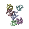 8b10C 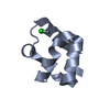 2f44S S: Starting model for refinement C: citing same article ( |
|---|---|
| Similar structure data |
-Links
 PDBj
PDBj
- Assembly
| Deposited unit | 
| ||||||||
|---|---|---|---|---|---|---|---|---|---|
| 1 |
| ||||||||
| Unit cell |
|
-Components
| #1: Protein | Mass: 8111.210 Da / Num. of mol.: 1 Source method: isolated from a genetically manipulated source Source: (gene. exp.) Homo sapiens (human) / Cell line: HEK 193T / Gene: SHANK2, CORTBP1, KIAA1022, PROSAP1 / Production host:  Escherichia coli (E. coli) / References: UniProt: Q9UPX8 Escherichia coli (E. coli) / References: UniProt: Q9UPX8 |
|---|---|
| #2: Chemical | ChemComp-FMT / Formic acid  Mass: 46.025 Da / Num. of mol.: 1 / Source method: obtained synthetically / Formula: CH2O2 Mass: 46.025 Da / Num. of mol.: 1 / Source method: obtained synthetically / Formula: CH2O2 |
| #3: Chemical | ChemComp-CL / Chloride  Mass: 35.453 Da / Num. of mol.: 1 / Source method: obtained synthetically / Formula: Cl Mass: 35.453 Da / Num. of mol.: 1 / Source method: obtained synthetically / Formula: Cl |
| #4: Chemical | ChemComp-ZN /   Mass: 65.409 Da / Num. of mol.: 1 / Source method: obtained synthetically / Formula: Zn / Feature type: SUBJECT OF INVESTIGATION Mass: 65.409 Da / Num. of mol.: 1 / Source method: obtained synthetically / Formula: Zn / Feature type: SUBJECT OF INVESTIGATION |
| #5: Water | ChemComp-HOH / Water Mass: 18.015 Da / Num. of mol.: 51 / Source method: isolated from a natural source / Formula: H2O Mass: 18.015 Da / Num. of mol.: 51 / Source method: isolated from a natural source / Formula: H2O |
| Has ligand of interest | Y |
-Experimental details
-Experiment
| Experiment | Method: X-RAY DIFFRACTION / Number of used crystals: 1 |
|---|
- Sample preparation
| Crystal | Density Matthews: 2.82 Å3/Da / Density % sol: 56.46 % |
|---|---|
| Crystal grow | Temperature: 292 K / Method: vapor diffusion, sitting drop / Details: 0.1M Bis Tris pH5.5, 0.3M of Magnesium formate |
-Data collection
| Diffraction | Mean temperature: 100 K / Serial crystal experiment: N |
|---|---|
| Diffraction source | Source: SYNCHROTRON / Site: PETRA III, EMBL c/o DESY / Beamline: P14 (MX2) / Wavelength: 0.9762 Å |
| Detector | Type: DECTRIS EIGER X 16M / Detector: PIXEL / Date: Mar 11, 2021 |
| Radiation | Protocol: SINGLE WAVELENGTH / Monochromatic (M) / Laue (L): M / Scattering type: x-ray |
| Radiation wavelength | Wavelength: 0.9762 Å / Relative weight: 1 |
| Reflection | Resolution: 2.1→49.8 Å / Num. obs: 5201 / % possible obs: 99.92 % / Redundancy: 19.8 % / CC1/2: 0.996 / Rmerge(I) obs: 0.2587 / Rpim(I) all: 0.05916 / Net I/σ(I): 10.07 |
| Reflection shell | Resolution: 2.1→2.16 Å / Redundancy: 19.7 % / Rmerge(I) obs: 1.42 / Mean I/σ(I) obs: 1.61 / Num. unique obs: 516 / CC1/2: 0.568 / Rpim(I) all: 0.3268 / % possible all: 100 |
- Processing
| Software |
| |||||||||||||||||||||||||||||||||||||||||||||||||||||||||||||||||||||||||||||||||||||||||||||||||||||||||||||||||||||||||||||||||||||||||||||||||||||||||||||||||||||||||||||||||||||||||||||||||||||||||||||||||||||||||||||||||||||||
|---|---|---|---|---|---|---|---|---|---|---|---|---|---|---|---|---|---|---|---|---|---|---|---|---|---|---|---|---|---|---|---|---|---|---|---|---|---|---|---|---|---|---|---|---|---|---|---|---|---|---|---|---|---|---|---|---|---|---|---|---|---|---|---|---|---|---|---|---|---|---|---|---|---|---|---|---|---|---|---|---|---|---|---|---|---|---|---|---|---|---|---|---|---|---|---|---|---|---|---|---|---|---|---|---|---|---|---|---|---|---|---|---|---|---|---|---|---|---|---|---|---|---|---|---|---|---|---|---|---|---|---|---|---|---|---|---|---|---|---|---|---|---|---|---|---|---|---|---|---|---|---|---|---|---|---|---|---|---|---|---|---|---|---|---|---|---|---|---|---|---|---|---|---|---|---|---|---|---|---|---|---|---|---|---|---|---|---|---|---|---|---|---|---|---|---|---|---|---|---|---|---|---|---|---|---|---|---|---|---|---|---|---|---|---|---|---|---|---|---|---|---|---|---|---|---|---|---|---|---|---|---|---|
| Refinement | Method to determine structure: MOLECULAR REPLACEMENT Starting model: 2F44 Resolution: 2.117→49.788 Å / Cor.coef. Fo:Fc: 0.957 / Cor.coef. Fo:Fc free: 0.926 / SU B: 8.048 / SU ML: 0.186 / Cross valid method: FREE R-VALUE / ESU R: 0.218 / ESU R Free: 0.196 Details: Hydrogens have been added in their riding positions
| |||||||||||||||||||||||||||||||||||||||||||||||||||||||||||||||||||||||||||||||||||||||||||||||||||||||||||||||||||||||||||||||||||||||||||||||||||||||||||||||||||||||||||||||||||||||||||||||||||||||||||||||||||||||||||||||||||||||
| Solvent computation | Ion probe radii: 0.8 Å / Shrinkage radii: 0.8 Å / VDW probe radii: 1.2 Å / Solvent model: MASK BULK SOLVENT | |||||||||||||||||||||||||||||||||||||||||||||||||||||||||||||||||||||||||||||||||||||||||||||||||||||||||||||||||||||||||||||||||||||||||||||||||||||||||||||||||||||||||||||||||||||||||||||||||||||||||||||||||||||||||||||||||||||||
| Displacement parameters | Biso mean: 38.747 Å2
| |||||||||||||||||||||||||||||||||||||||||||||||||||||||||||||||||||||||||||||||||||||||||||||||||||||||||||||||||||||||||||||||||||||||||||||||||||||||||||||||||||||||||||||||||||||||||||||||||||||||||||||||||||||||||||||||||||||||
| Refinement step | Cycle: LAST / Resolution: 2.117→49.788 Å
| |||||||||||||||||||||||||||||||||||||||||||||||||||||||||||||||||||||||||||||||||||||||||||||||||||||||||||||||||||||||||||||||||||||||||||||||||||||||||||||||||||||||||||||||||||||||||||||||||||||||||||||||||||||||||||||||||||||||
| Refine LS restraints |
| |||||||||||||||||||||||||||||||||||||||||||||||||||||||||||||||||||||||||||||||||||||||||||||||||||||||||||||||||||||||||||||||||||||||||||||||||||||||||||||||||||||||||||||||||||||||||||||||||||||||||||||||||||||||||||||||||||||||
| LS refinement shell | Refine-ID: X-RAY DIFFRACTION / Total num. of bins used: 20
|
